H J Odendaal, I C Crockart, C Du Plessis, L Brink, C A Groenewald
{"title":"Accelerations of the Fetal Heart Rate in the Screening for Fetal Growth Restriction at 34-38 Week's Gestation.","authors":"H J Odendaal, I C Crockart, C Du Plessis, L Brink, C A Groenewald","doi":"","DOIUrl":null,"url":null,"abstract":"<p><strong>Objectives: </strong>To use machine learning to determine what information on Doppler velocimetry and maternal and fetal heart rates, collected at 20-24 weeks gestation, correlates best with fetal growth restriction according to the estimated fetal weight at 34-38 weeks.</p><p><strong>Study design: </strong>Data of 4496 pregnant women, collected prospectively for the Safe Passage Study, from August 2007 to August 2016, were used for the present analysis. Doppler flow velocity of the uterine, umbilical, and middle cerebral arteries and transabdominally recorded maternal and fetal ECGs were collected at 20-24 weeks gestation and fetal biometry collected at 34-38 weeks from which the estimated fetal weight was calculated. Fetal growth restriction was defined as an estimated fetal weight below the 10th centile. Accelerations and decelerations of the fetal and maternal heart rates were quantified as gained or lost beats per hour of recording respectively. Machine learning with receiver operative characteristic curves were then used to determine which model gives the best performance.</p><p><strong>Results: </strong>The final model performed exceptionally well across all evaluation metrics, particularly so for the Stochastic Gradient Descent method: achieving a 93% average for Classification Accuracy, Recall, Precision and F1-Score to identify the fetus with an estimated weight below the 10th percentile at 34-38 weeks. Ranking determined that the most important standard feature was the umbilical artery pulsatility index. However, the excellent overall accuracy is likely due to the value added by the pre-processed features regarding fetal gained beats and accelerations.</p><p><strong>Conclusion: </strong>Fetal movements, as characterized by gained beats as early as 20-24 weeks gestation, contribute to the value of the flow velocimetry of the umbilical artery at 34-38 weeks in identifying the growth restricted fetus.</p>","PeriodicalId":87261,"journal":{"name":"Global journal of pediatrics & neonatal care","volume":"3 5","pages":""},"PeriodicalIF":0.0000,"publicationDate":"2021-01-01","publicationTypes":"Journal Article","fieldsOfStudy":null,"isOpenAccess":false,"openAccessPdf":"https://www.ncbi.nlm.nih.gov/pmc/articles/PMC8607280/pdf/","citationCount":"0","resultStr":null,"platform":"Semanticscholar","paperid":null,"PeriodicalName":"Global journal of pediatrics & neonatal care","FirstCategoryId":"1085","ListUrlMain":"","RegionNum":0,"RegionCategory":null,"ArticlePicture":[],"TitleCN":null,"AbstractTextCN":null,"PMCID":null,"EPubDate":"2021/10/30 0:00:00","PubModel":"Epub","JCR":"","JCRName":"","Score":null,"Total":0}
引用次数: 0
Abstract
Objectives: To use machine learning to determine what information on Doppler velocimetry and maternal and fetal heart rates, collected at 20-24 weeks gestation, correlates best with fetal growth restriction according to the estimated fetal weight at 34-38 weeks.
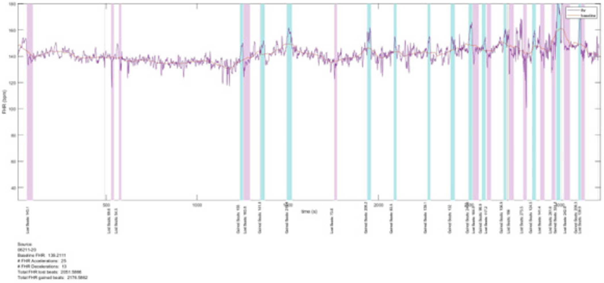
Study design: Data of 4496 pregnant women, collected prospectively for the Safe Passage Study, from August 2007 to August 2016, were used for the present analysis. Doppler flow velocity of the uterine, umbilical, and middle cerebral arteries and transabdominally recorded maternal and fetal ECGs were collected at 20-24 weeks gestation and fetal biometry collected at 34-38 weeks from which the estimated fetal weight was calculated. Fetal growth restriction was defined as an estimated fetal weight below the 10th centile. Accelerations and decelerations of the fetal and maternal heart rates were quantified as gained or lost beats per hour of recording respectively. Machine learning with receiver operative characteristic curves were then used to determine which model gives the best performance.
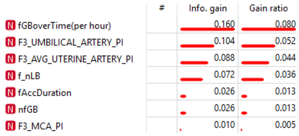
Results: The final model performed exceptionally well across all evaluation metrics, particularly so for the Stochastic Gradient Descent method: achieving a 93% average for Classification Accuracy, Recall, Precision and F1-Score to identify the fetus with an estimated weight below the 10th percentile at 34-38 weeks. Ranking determined that the most important standard feature was the umbilical artery pulsatility index. However, the excellent overall accuracy is likely due to the value added by the pre-processed features regarding fetal gained beats and accelerations.
Conclusion: Fetal movements, as characterized by gained beats as early as 20-24 weeks gestation, contribute to the value of the flow velocimetry of the umbilical artery at 34-38 weeks in identifying the growth restricted fetus.

 求助内容:
求助内容: 应助结果提醒方式:


