Eero Rissanen, Kelsey Carter, Steven Cicero, John Ficke, Marie Kijewski, Mi-Ae Park, Joseph Kijewski, Emily Stern, Tanuja Chitnis, David Silbersweig, Howard L Weiner, Chun K Kim, Jennifer Lyons, Joshua P Klein, Shamik Bhattacharyya, Tarun Singhal
{"title":"Cortical and Subcortical Dysmetabolism Are Dynamic Markers of Clinical Disability and Course in Anti-LGI1 Encephalitis.","authors":"Eero Rissanen, Kelsey Carter, Steven Cicero, John Ficke, Marie Kijewski, Mi-Ae Park, Joseph Kijewski, Emily Stern, Tanuja Chitnis, David Silbersweig, Howard L Weiner, Chun K Kim, Jennifer Lyons, Joshua P Klein, Shamik Bhattacharyya, Tarun Singhal","doi":"10.1212/NXI.0000000000001136","DOIUrl":null,"url":null,"abstract":"<p><strong>Background and objectives: </strong>This [<sup>18</sup>F]fluorodeoxyglucose (FDG) PET study evaluates the accuracy of semiquantitative measurement of putaminal hypermetabolism in identifying anti-leucine-rich, glioma-inactivated-1 (LGI1) protein autoimmune encephalitis (AE). In addition, the extent of brain dysmetabolism, their association with clinical outcomes, and longitudinal metabolic changes after immunotherapy in LGI1-AE are examined.</p><p><strong>Methods: </strong>FDG-PET scans from 49 age-matched and sex-matched subjects (13 in LGI1-AE group, 15 in non-LGI1-AE group, 11 with Alzheimer disease [AD], and 10 negative controls [NCs]) and follow-up scans from 8 patients with LGI1 AE on a median 6 months after immunotherapy were analyzed. Putaminal standardized uptake value ratios (SUVRs) normalized to global brain (P-SUVRg), thalamus (P/Th), and midbrain (P/Mi) were evaluated for diagnostic accuracy. SUVRg was applied for all other analyses.</p><p><strong>Results: </strong>P-SUVRg, P/Th, and P/Mi were higher in LGI1-AE group than in non-LGI1-AE group, AD group, and NCs (all <i>p</i> < 0.05). P/Mi and P-SUVRg differentiated LGI1-AE group robustly from other groups (areas under the curve 0.84-0.99). Mediotemporal lobe (MTL) SUVRg was increased in both LGI1-AE and non-LGI1-AE groups when compared with NCs (both <i>p</i> < 0.05). SUVRg was decreased in several frontoparietal regions and increased in pallidum, caudate, pons, olfactory, and inferior occipital gyrus in LGI1-AE group when compared with that in NCs (all <i>p</i> < 0.05). In LGI1-AE group, both MTL and putaminal hypermetabolism were reduced after immunotherapy. Normalization of regional cortical dysmetabolism associated with clinical improvement at the 6- and 20-month follow-up.</p><p><strong>Discussion: </strong>Semiquantitative measurement of putaminal hypermetabolism with FDG-PET may be used to distinguish LGI1-AE from other pathologies. Metabolic abnormalities in LGI1-AE extend beyond putamen and MTL into other subcortical and cortical regions. FDG-PET may be used in evaluating disease evolution in LGI1-AE.</p><p><strong>Classification of evidence: </strong>This study provides Class II evidence that semiquantitative measures of putaminal metabolism on PET can differentiate patients with LGI1-AE from patients without LGI1-AE, patients with AD, or NCs.</p>","PeriodicalId":520720,"journal":{"name":"Neurology(R) neuroimmunology & neuroinflammation","volume":" ","pages":""},"PeriodicalIF":7.5000,"publicationDate":"2022-01-28","publicationTypes":"Journal Article","fieldsOfStudy":null,"isOpenAccess":false,"openAccessPdf":"https://ftp.ncbi.nlm.nih.gov/pub/pmc/oa_pdf/eb/c3/NEURIMMINFL2021039229.PMC8802686.pdf","citationCount":"10","resultStr":null,"platform":"Semanticscholar","paperid":null,"PeriodicalName":"Neurology(R) neuroimmunology & neuroinflammation","FirstCategoryId":"3","ListUrlMain":"https://doi.org/10.1212/NXI.0000000000001136","RegionNum":0,"RegionCategory":null,"ArticlePicture":[],"TitleCN":null,"AbstractTextCN":null,"PMCID":null,"EPubDate":"2022/3/1 0:00:00","PubModel":"Print","JCR":"","JCRName":"","Score":null,"Total":0}
引用次数: 10
Abstract
Background and objectives: This [18F]fluorodeoxyglucose (FDG) PET study evaluates the accuracy of semiquantitative measurement of putaminal hypermetabolism in identifying anti-leucine-rich, glioma-inactivated-1 (LGI1) protein autoimmune encephalitis (AE). In addition, the extent of brain dysmetabolism, their association with clinical outcomes, and longitudinal metabolic changes after immunotherapy in LGI1-AE are examined.
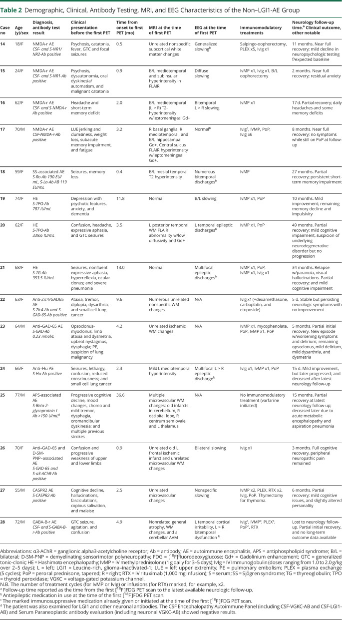
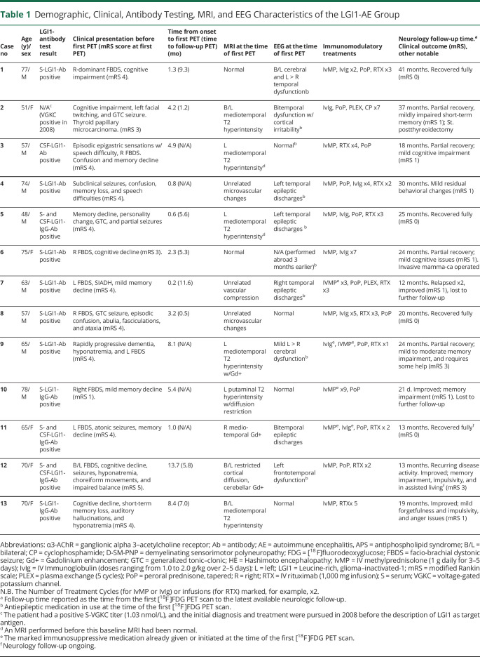
Methods: FDG-PET scans from 49 age-matched and sex-matched subjects (13 in LGI1-AE group, 15 in non-LGI1-AE group, 11 with Alzheimer disease [AD], and 10 negative controls [NCs]) and follow-up scans from 8 patients with LGI1 AE on a median 6 months after immunotherapy were analyzed. Putaminal standardized uptake value ratios (SUVRs) normalized to global brain (P-SUVRg), thalamus (P/Th), and midbrain (P/Mi) were evaluated for diagnostic accuracy. SUVRg was applied for all other analyses.
Results: P-SUVRg, P/Th, and P/Mi were higher in LGI1-AE group than in non-LGI1-AE group, AD group, and NCs (all p < 0.05). P/Mi and P-SUVRg differentiated LGI1-AE group robustly from other groups (areas under the curve 0.84-0.99). Mediotemporal lobe (MTL) SUVRg was increased in both LGI1-AE and non-LGI1-AE groups when compared with NCs (both p < 0.05). SUVRg was decreased in several frontoparietal regions and increased in pallidum, caudate, pons, olfactory, and inferior occipital gyrus in LGI1-AE group when compared with that in NCs (all p < 0.05). In LGI1-AE group, both MTL and putaminal hypermetabolism were reduced after immunotherapy. Normalization of regional cortical dysmetabolism associated with clinical improvement at the 6- and 20-month follow-up.
Discussion: Semiquantitative measurement of putaminal hypermetabolism with FDG-PET may be used to distinguish LGI1-AE from other pathologies. Metabolic abnormalities in LGI1-AE extend beyond putamen and MTL into other subcortical and cortical regions. FDG-PET may be used in evaluating disease evolution in LGI1-AE.
Classification of evidence: This study provides Class II evidence that semiquantitative measures of putaminal metabolism on PET can differentiate patients with LGI1-AE from patients without LGI1-AE, patients with AD, or NCs.

 求助内容:
求助内容: 应助结果提醒方式:


