{"title":"Optic Nerve Lesion Length at the Acute Phase of Optic Neuritis Is Predictive of Retinal Neuronal Loss.","authors":"Mickael Denis, Jean-Philippe Woillez, Vasily M Smirnov, Elodie Drumez, Julien Lannoy, Julie Boucher, Mickael Zedet, Jean-Pierre Pruvo, Julien Labreuche, Helene Zephir, Xavier Leclerc, Olivier Outteryck","doi":"10.1212/NXI.0000000000001135","DOIUrl":null,"url":null,"abstract":"<p><strong>Background and objectives: </strong>Acute optic neuritis (ON) is a classical presenting symptom of multiple sclerosis (MS), neuromyelitis optica spectrum disorders (NMOSD), and anti-MOG-associated disorders. The resulting visual impairment is variable and can be severe. Clinicians are in need of predictive biomarkers to optimize the management of acute ON. In this longitudinal study (IRMANO, NCT03651662), we evaluated the ability of optic nerve lesion length measured on MRI at the acute phase of ON to predict retinal neuro-axonal loss and visual impairment at a chronic stage.</p><p><strong>Methods: </strong>We conducted a longitudinal study (IRMANO, NCT03651662) of patients who presented a clinical episode of ON (≤8 weeks). All patients underwent a retinal optical coherence tomography (OCT) and a brain/optic nerve MRI, including 3D double-inversion recovery (DIR) sequence at the acute phase of ON and 12 months later. Primary outcomes were optic nerve DIR hypersignal lesion length, macular ganglion cell-inner plexiform layer (GCIPL) volume measured on OCT, and low-contrast monocular visual acuity (LCMVA).</p><p><strong>Results: </strong>The study group included 51 patients (33 women, mean age of 32.4 years ± 7.9). We recruited patients with a clinically isolated syndrome (n = 20), a relapsing-remitting MS (n = 23), an isolated ON (n = 6), and a first clinical episode of NMOSD (n = 2). Optic nerve DIR hypersignal was observed in all but 1 symptomatic optic nerves. At inclusion, the mean optic nerve lesion length (in mm) was 12.35 ± 5.98. The mean GCIPL volume (in mm<sup>3</sup>) significantly decreased between inclusion (1.90 ± 0.18) and M<sub>12</sub> (1.67 ± 0.21; <i>p</i> < 0.0001). Optic nerve lesion length at inclusion was significantly associated with GCIPL thinning (estimate ± SD; -0.012 ± 0.004; <i>p</i> = 0.0016) and LCMVA at M<sub>12</sub> (0.016 ± 0.003; <i>p</i> < 0.001). Optic nerve lesion length significantly increased at M<sub>12</sub> (15.76 ± 8.70; <i>p</i> = 0.0007). The increase in optic nerve lesion length was significantly associated with the GCIPL thinning between inclusion and M<sub>12</sub> (-0.012 ± 0.003; <i>p</i> = 0.0011).</p><p><strong>Discussion: </strong>At the acute phase of ON, optic nerve lesion length is an imaging biomarker predictive of retinal neuro-axonal loss and chronic visual impairment, which can help to stratify future therapeutic strategies in acute ON.</p><p><strong>Classification of evidence: </strong>This study provides Class I evidence that optic nerve lesion length measured on MRI during the acute phase of a first episode of ON is associated with long-term retinal neuro-axonal loss and visual impairment.</p>","PeriodicalId":520720,"journal":{"name":"Neurology(R) neuroimmunology & neuroinflammation","volume":" ","pages":""},"PeriodicalIF":7.5000,"publicationDate":"2022-01-28","publicationTypes":"Journal Article","fieldsOfStudy":null,"isOpenAccess":false,"openAccessPdf":"https://ftp.ncbi.nlm.nih.gov/pub/pmc/oa_pdf/63/d0/NEURIMMINFL2021039451.PMC8802684.pdf","citationCount":"13","resultStr":null,"platform":"Semanticscholar","paperid":null,"PeriodicalName":"Neurology(R) neuroimmunology & neuroinflammation","FirstCategoryId":"3","ListUrlMain":"https://doi.org/10.1212/NXI.0000000000001135","RegionNum":0,"RegionCategory":null,"ArticlePicture":[],"TitleCN":null,"AbstractTextCN":null,"PMCID":null,"EPubDate":"2022/3/1 0:00:00","PubModel":"Print","JCR":"","JCRName":"","Score":null,"Total":0}
引用次数: 13
Abstract
Background and objectives: Acute optic neuritis (ON) is a classical presenting symptom of multiple sclerosis (MS), neuromyelitis optica spectrum disorders (NMOSD), and anti-MOG-associated disorders. The resulting visual impairment is variable and can be severe. Clinicians are in need of predictive biomarkers to optimize the management of acute ON. In this longitudinal study (IRMANO, NCT03651662), we evaluated the ability of optic nerve lesion length measured on MRI at the acute phase of ON to predict retinal neuro-axonal loss and visual impairment at a chronic stage.
Methods: We conducted a longitudinal study (IRMANO, NCT03651662) of patients who presented a clinical episode of ON (≤8 weeks). All patients underwent a retinal optical coherence tomography (OCT) and a brain/optic nerve MRI, including 3D double-inversion recovery (DIR) sequence at the acute phase of ON and 12 months later. Primary outcomes were optic nerve DIR hypersignal lesion length, macular ganglion cell-inner plexiform layer (GCIPL) volume measured on OCT, and low-contrast monocular visual acuity (LCMVA).
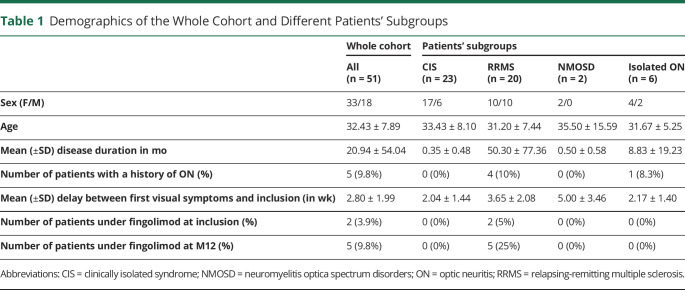
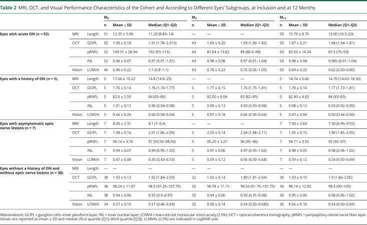
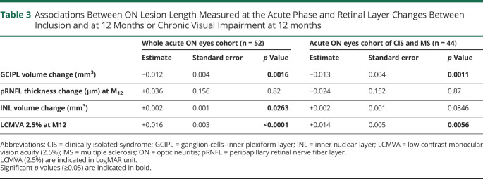
Results: The study group included 51 patients (33 women, mean age of 32.4 years ± 7.9). We recruited patients with a clinically isolated syndrome (n = 20), a relapsing-remitting MS (n = 23), an isolated ON (n = 6), and a first clinical episode of NMOSD (n = 2). Optic nerve DIR hypersignal was observed in all but 1 symptomatic optic nerves. At inclusion, the mean optic nerve lesion length (in mm) was 12.35 ± 5.98. The mean GCIPL volume (in mm3) significantly decreased between inclusion (1.90 ± 0.18) and M12 (1.67 ± 0.21; p < 0.0001). Optic nerve lesion length at inclusion was significantly associated with GCIPL thinning (estimate ± SD; -0.012 ± 0.004; p = 0.0016) and LCMVA at M12 (0.016 ± 0.003; p < 0.001). Optic nerve lesion length significantly increased at M12 (15.76 ± 8.70; p = 0.0007). The increase in optic nerve lesion length was significantly associated with the GCIPL thinning between inclusion and M12 (-0.012 ± 0.003; p = 0.0011).
Discussion: At the acute phase of ON, optic nerve lesion length is an imaging biomarker predictive of retinal neuro-axonal loss and chronic visual impairment, which can help to stratify future therapeutic strategies in acute ON.
Classification of evidence: This study provides Class I evidence that optic nerve lesion length measured on MRI during the acute phase of a first episode of ON is associated with long-term retinal neuro-axonal loss and visual impairment.

 求助内容:
求助内容: 应助结果提醒方式:


