Rouholla Bagheri, Jalal Haghighat Monfared, Mohammad Reza Montazeriyoun
{"title":"Brain Tumor Segmentation Using Graph Coloring Approach in Magnetic Resonance Images.","authors":"Rouholla Bagheri, Jalal Haghighat Monfared, Mohammad Reza Montazeriyoun","doi":"10.4103/jmss.JMSS_43_20","DOIUrl":null,"url":null,"abstract":"<p><p>It is important to have an accurate and reliable brain tumor segmentation for cancer diagnosis and treatment planning. There are few unsupervised approaches for brain tumor segmentation. In this paper, a new unsupervised approach based on graph coloring for brain tumor segmentation is introduced. In this study, a graph coloring approach is used for brain tumor segmentation. For this aim, each pixel of brain image assumed as a node of graph and difference between brightness of a couple of pixels considered as edge. This method was applied on T1-enhanced magnetic resonance images of low-grade and high-grade patients. Since a rigid graph was needed for graph coloring, edges must be divided into existing or nonexisting edge using a threshold. The value of this threshold has affected the accuracy of image segmentation, so the choice of the optimal threshold was important. The optimal value for this threshold was 0.42 of maximum value of difference of brightness between pixels that caused the 83.62% of correlation accuracy. The results showed that graph coloring approach can be a reliable unsupervised approach for brain tumor segmentation. This approach, as an unsupervised approach, shows better accuracy in comparison with neural networks and neuro-fuzzy networks. However, as a limitation, the accuracy of this approach is dependent on the threshold of edges.</p>","PeriodicalId":37680,"journal":{"name":"Journal of Medical Signals & Sensors","volume":"11 4","pages":"285-290"},"PeriodicalIF":1.1000,"publicationDate":"2021-10-20","publicationTypes":"Journal Article","fieldsOfStudy":null,"isOpenAccess":false,"openAccessPdf":"https://ftp.ncbi.nlm.nih.gov/pub/pmc/oa_pdf/86/ec/JMSS-11-285.PMC8588878.pdf","citationCount":"0","resultStr":null,"platform":"Semanticscholar","paperid":null,"PeriodicalName":"Journal of Medical Signals & Sensors","FirstCategoryId":"1085","ListUrlMain":"https://doi.org/10.4103/jmss.JMSS_43_20","RegionNum":0,"RegionCategory":null,"ArticlePicture":[],"TitleCN":null,"AbstractTextCN":null,"PMCID":null,"EPubDate":"2021/10/1 0:00:00","PubModel":"eCollection","JCR":"Q4","JCRName":"ENGINEERING, BIOMEDICAL","Score":null,"Total":0}
引用次数: 0
Abstract
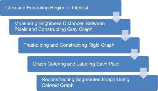
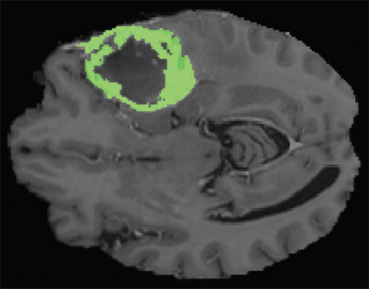
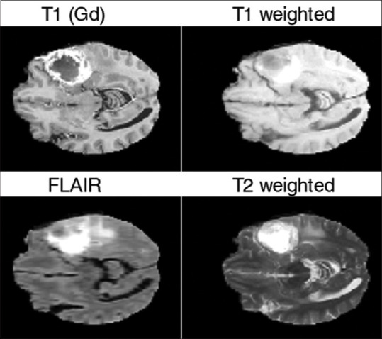
It is important to have an accurate and reliable brain tumor segmentation for cancer diagnosis and treatment planning. There are few unsupervised approaches for brain tumor segmentation. In this paper, a new unsupervised approach based on graph coloring for brain tumor segmentation is introduced. In this study, a graph coloring approach is used for brain tumor segmentation. For this aim, each pixel of brain image assumed as a node of graph and difference between brightness of a couple of pixels considered as edge. This method was applied on T1-enhanced magnetic resonance images of low-grade and high-grade patients. Since a rigid graph was needed for graph coloring, edges must be divided into existing or nonexisting edge using a threshold. The value of this threshold has affected the accuracy of image segmentation, so the choice of the optimal threshold was important. The optimal value for this threshold was 0.42 of maximum value of difference of brightness between pixels that caused the 83.62% of correlation accuracy. The results showed that graph coloring approach can be a reliable unsupervised approach for brain tumor segmentation. This approach, as an unsupervised approach, shows better accuracy in comparison with neural networks and neuro-fuzzy networks. However, as a limitation, the accuracy of this approach is dependent on the threshold of edges.
期刊介绍:
JMSS is an interdisciplinary journal that incorporates all aspects of the biomedical engineering including bioelectrics, bioinformatics, medical physics, health technology assessment, etc. Subject areas covered by the journal include: - Bioelectric: Bioinstruments Biosensors Modeling Biomedical signal processing Medical image analysis and processing Medical imaging devices Control of biological systems Neuromuscular systems Cognitive sciences Telemedicine Robotic Medical ultrasonography Bioelectromagnetics Electrophysiology Cell tracking - Bioinformatics and medical informatics: Analysis of biological data Data mining Stochastic modeling Computational genomics Artificial intelligence & fuzzy Applications Medical softwares Bioalgorithms Electronic health - Biophysics and medical physics: Computed tomography Radiation therapy Laser therapy - Education in biomedical engineering - Health technology assessment - Standard in biomedical engineering.

 求助内容:
求助内容: 应助结果提醒方式:


