[Spontaneous expulsion of a mesenchymal colonic tumor via the rectum].
4区 医学
Q3 Medicine
引用次数: 0
Abstract
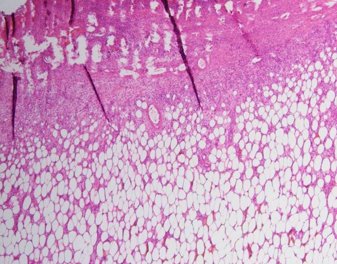
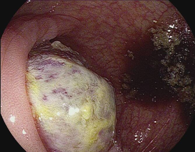
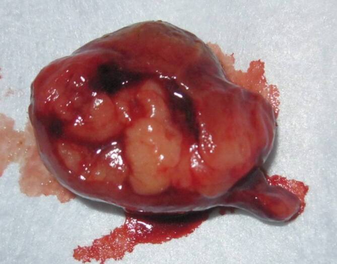
A 61-year-old male patient underwent a colonoscopy for cramp-like upper abdominal pain of 3 weeks duration. An endoscopically irresectable ulcerated mass was seen in the transverse colon. The patient spontaneously excreted in the feces a tumor node measuring 4.1 × 3.5 × 2.8 cm with the histological features of a submucosal lipoma 4 days after the colonoscopy. A benign lipoma confined to the submucosa was operatively confirmed. It is extremely rare for a tumor node to be shed in feces. If the benign nature of the entire lesion is doubtful, standard oncological procedures are advocated.
[结肠间充质肿瘤经直肠自发排出]。
61岁男性患者因抽筋样上腹部疼痛3周行结肠镜检查。内窥镜下发现横结肠不可切除的溃疡肿块。患者于结肠镜检查4天后,粪便中自发排出肿瘤结1个,尺寸为4.1 × 3.5 × 2.8 cm,组织学表现为黏膜下脂肪瘤。手术证实为局限于粘膜下层的良性脂肪瘤。肿瘤结随粪便排出是极为罕见的。如果整个病变的良性性质是可疑的,则提倡标准的肿瘤治疗程序。
本文章由计算机程序翻译,如有差异,请以英文原文为准。
求助全文
约1分钟内获得全文
求助全文
来源期刊
Internist
医学-医学:内科
CiteScore
1.20
自引率
0.00%
发文量
139
审稿时长
4-8 weeks
期刊介绍:
Der Internist is an internationally respected journal dealing with all aspects of internal medicine. The journal serves both the scientific exchange and the continuing education of internists working in practical or clinical environments as well as of general practitioners who are particularly interested in internal medicine. The focus is on the topics of prevention, diagnostic approaches, management of complications, and current therapy strategies.
Comprehensive reviews on a specific topical issue focus on providing evidenced based information on diagnostics and therapy.
Case reports feature interesting cases and aim at optimizing diagnostic and therapeutic strategies.
Review articles under the rubric "Continuing Medical Education" present verified results of scientific research and their integration into daily practice.
 求助内容:
求助内容: 应助结果提醒方式:


