Dina Mönch, Jana Koch, Annika Maaß, Nicole Janssen, Thomas Mürdter, Philipp Renner, Petra Fallier-Becker, Wiebke Solaß, Matthias Schwab, Marc-H Dahlke, Hans J Schlitt, Tobias Leibold
{"title":"A human <i>ex vivo</i> coculture model to investigate peritoneal metastasis and innovative treatment options.","authors":"Dina Mönch, Jana Koch, Annika Maaß, Nicole Janssen, Thomas Mürdter, Philipp Renner, Petra Fallier-Becker, Wiebke Solaß, Matthias Schwab, Marc-H Dahlke, Hans J Schlitt, Tobias Leibold","doi":"10.1515/pp-2021-0128","DOIUrl":null,"url":null,"abstract":"<p><strong>Objectives: </strong>Peritoneal metastasis (PM) is commonly observed in patients with colorectal cancer (CRC). The outcome of these patients is poor, with an average survival of only six months without therapy, which requires a better understanding of PM biology and new treatment strategies.</p><p><strong>Methods: </strong>We established and characterized a human <i>ex vivo</i> peritoneal model to investigate the mechanisms of peritoneal seeding and possible treatment options. For this, CRC cell lines and patient-derived tumor organoids were cultured together with human peritoneum to investigate the invasion of malignant cells and the effects of local chemotherapy.</p><p><strong>Results: </strong>Fresh human peritoneum was cultured for up to three weeks in a stainless steel ring system, allowing for survival of all peritoneal structures. Peritoneal cell survival was documented by light microscopy and immunohistochemical staining. Further, immunohistological characterization of the tissue revealed CD3-positive T-lymphocytes and vimentin-positive fibroblasts within the peritoneum. In addition, extracellular matrix components (collagens, matrix metalloproteinases) were localized within the tissue. Coculture with CRC cell lines and patient-derived CRC organoids revealed that cancer cells grew on the peritoneum and migrated into the tissue. Coculture with CRC cells confirmed that hyperthermal treatment at 41 °C for 90 min significantly enhanced the intracellular entry of doxorubicin. Moreover, treatment with mitomycin C under hyperthermic conditions significantly reduced the amount of cancer cells within the peritoneum.</p><p><strong>Conclusions: </strong>This human <i>ex vivo</i> peritoneal model provides a stringent and clinically relevant platform for the investigation of PM and for further elucidation of possible treatment options.</p>","PeriodicalId":20231,"journal":{"name":"Pleura and Peritoneum","volume":"6 3","pages":"121-129"},"PeriodicalIF":2.4000,"publicationDate":"2021-07-27","publicationTypes":"Journal Article","fieldsOfStudy":null,"isOpenAccess":false,"openAccessPdf":"https://sci-hub-pdf.com/10.1515/pp-2021-0128","citationCount":"1","resultStr":null,"platform":"Semanticscholar","paperid":null,"PeriodicalName":"Pleura and Peritoneum","FirstCategoryId":"1085","ListUrlMain":"https://doi.org/10.1515/pp-2021-0128","RegionNum":0,"RegionCategory":null,"ArticlePicture":[],"TitleCN":null,"AbstractTextCN":null,"PMCID":null,"EPubDate":"2021/9/1 0:00:00","PubModel":"eCollection","JCR":"Q4","JCRName":"ONCOLOGY","Score":null,"Total":0}
引用次数: 1
Abstract
Objectives: Peritoneal metastasis (PM) is commonly observed in patients with colorectal cancer (CRC). The outcome of these patients is poor, with an average survival of only six months without therapy, which requires a better understanding of PM biology and new treatment strategies.
Methods: We established and characterized a human ex vivo peritoneal model to investigate the mechanisms of peritoneal seeding and possible treatment options. For this, CRC cell lines and patient-derived tumor organoids were cultured together with human peritoneum to investigate the invasion of malignant cells and the effects of local chemotherapy.
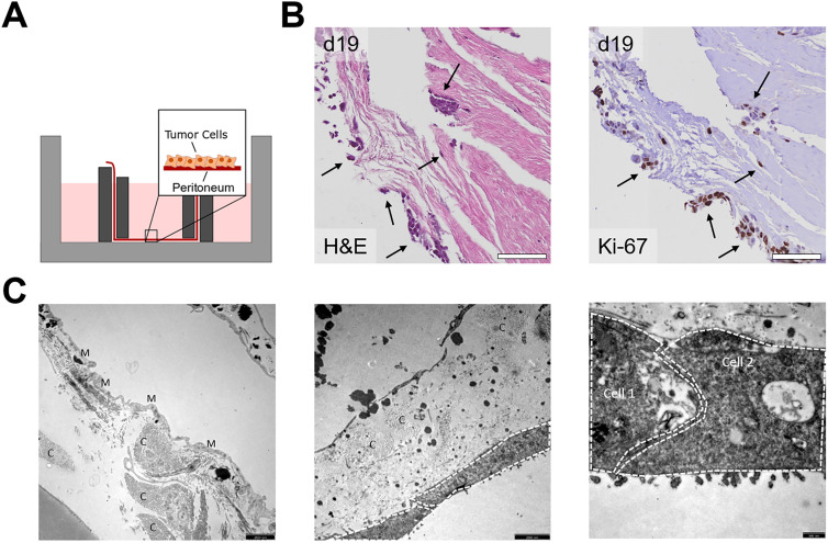
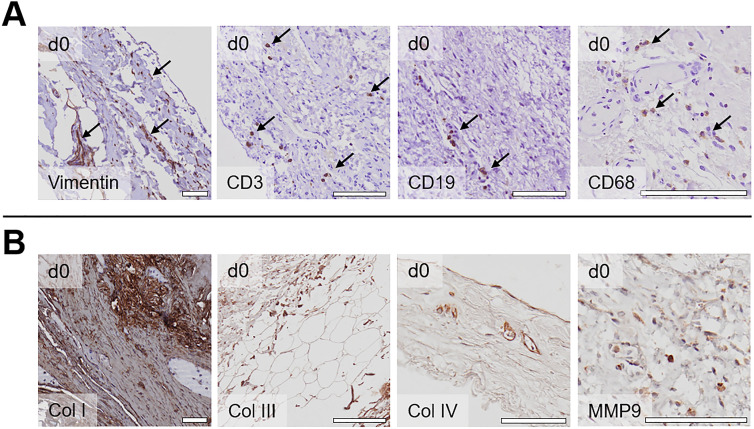
Results: Fresh human peritoneum was cultured for up to three weeks in a stainless steel ring system, allowing for survival of all peritoneal structures. Peritoneal cell survival was documented by light microscopy and immunohistochemical staining. Further, immunohistological characterization of the tissue revealed CD3-positive T-lymphocytes and vimentin-positive fibroblasts within the peritoneum. In addition, extracellular matrix components (collagens, matrix metalloproteinases) were localized within the tissue. Coculture with CRC cell lines and patient-derived CRC organoids revealed that cancer cells grew on the peritoneum and migrated into the tissue. Coculture with CRC cells confirmed that hyperthermal treatment at 41 °C for 90 min significantly enhanced the intracellular entry of doxorubicin. Moreover, treatment with mitomycin C under hyperthermic conditions significantly reduced the amount of cancer cells within the peritoneum.
Conclusions: This human ex vivo peritoneal model provides a stringent and clinically relevant platform for the investigation of PM and for further elucidation of possible treatment options.


 求助内容:
求助内容: 应助结果提醒方式:


