Elisa Penna, Christopher L Cunningham, Stephanie Saylor, Anna Kreutz, Alice F Tarantal, Verónica Martínez-Cerdeño, Stephen C Noctor
{"title":"Greater Number of Microglia in Telencephalic Proliferative Zones of Human and Nonhuman Primate Compared with Other Vertebrate Species.","authors":"Elisa Penna, Christopher L Cunningham, Stephanie Saylor, Anna Kreutz, Alice F Tarantal, Verónica Martínez-Cerdeño, Stephen C Noctor","doi":"10.1093/texcom/tgab053","DOIUrl":null,"url":null,"abstract":"<p><p>Microglial cells, the innate immune cells of the brain, are derived from yolk sac precursor cells, begin to colonize the telencephalon at the onset of cortical neurogenesis, and occupy specific layers including the telencephalic proliferative zones. Microglia are an intrinsic component of cortical germinal zones, establish extensive contacts with neural precursor cells (NPCs) and developing cortical vessels, and regulate the size of the NPC pool through mechanisms that include phagocytosis. Microglia exhibit notable differences in number and distribution in the prenatal neocortex between rat and old world nonhuman primate telencephalon, suggesting that microglia possess distinct properties across vertebrate species. To begin addressing this subject, we quantified the number of microglia and NPCs in proliferative zones of the fetal human, rhesus monkey, ferret, and rat, and the prehatch chick and turtle telencephalon. We show that the ratio of NPCs to microglia varies significantly across species. Few microglia populate the prehatch chick telencephalon, but the number of microglia approaches that of NPCs in fetal human and nonhuman primate telencephalon. These data demonstrate that microglia are in a position to perform important functions in a number of vertebrate species but more heavily colonize proliferative zones of fetal human and rhesus monkey telencephalon.</p>","PeriodicalId":72551,"journal":{"name":"Cerebral cortex communications","volume":" ","pages":"tgab053"},"PeriodicalIF":0.0000,"publicationDate":"2021-09-06","publicationTypes":"Journal Article","fieldsOfStudy":null,"isOpenAccess":false,"openAccessPdf":"https://www.ncbi.nlm.nih.gov/pmc/articles/PMC8501267/pdf/","citationCount":"0","resultStr":null,"platform":"Semanticscholar","paperid":null,"PeriodicalName":"Cerebral cortex communications","FirstCategoryId":"1085","ListUrlMain":"https://doi.org/10.1093/texcom/tgab053","RegionNum":0,"RegionCategory":null,"ArticlePicture":[],"TitleCN":null,"AbstractTextCN":null,"PMCID":null,"EPubDate":"2021/1/1 0:00:00","PubModel":"eCollection","JCR":"","JCRName":"","Score":null,"Total":0}
引用次数: 0
Abstract
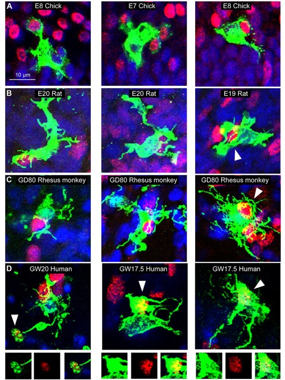
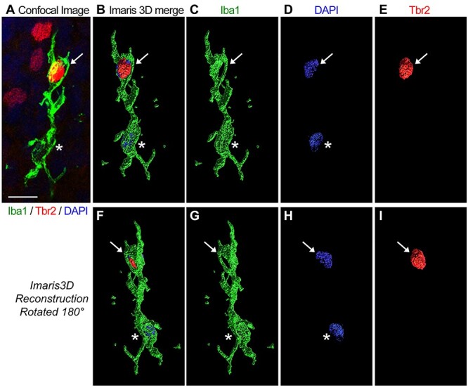
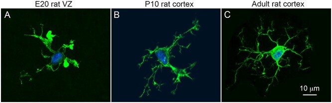
Microglial cells, the innate immune cells of the brain, are derived from yolk sac precursor cells, begin to colonize the telencephalon at the onset of cortical neurogenesis, and occupy specific layers including the telencephalic proliferative zones. Microglia are an intrinsic component of cortical germinal zones, establish extensive contacts with neural precursor cells (NPCs) and developing cortical vessels, and regulate the size of the NPC pool through mechanisms that include phagocytosis. Microglia exhibit notable differences in number and distribution in the prenatal neocortex between rat and old world nonhuman primate telencephalon, suggesting that microglia possess distinct properties across vertebrate species. To begin addressing this subject, we quantified the number of microglia and NPCs in proliferative zones of the fetal human, rhesus monkey, ferret, and rat, and the prehatch chick and turtle telencephalon. We show that the ratio of NPCs to microglia varies significantly across species. Few microglia populate the prehatch chick telencephalon, but the number of microglia approaches that of NPCs in fetal human and nonhuman primate telencephalon. These data demonstrate that microglia are in a position to perform important functions in a number of vertebrate species but more heavily colonize proliferative zones of fetal human and rhesus monkey telencephalon.

 求助内容:
求助内容: 应助结果提醒方式:


