Alexander L Lazarides, Harrison R Ferlauto, Zachary D C Burke, Anthony M Griffin, Bruce D Leckey, Nicholas M Bernthal, Jay S Wunder, Peter C Ferguson, Julia D Visgauss, Brian E Brigman, William C Eward
{"title":"The Utility of Chest Imaging for Surveillance of Atypical Lipomatous Tumors.","authors":"Alexander L Lazarides, Harrison R Ferlauto, Zachary D C Burke, Anthony M Griffin, Bruce D Leckey, Nicholas M Bernthal, Jay S Wunder, Peter C Ferguson, Julia D Visgauss, Brian E Brigman, William C Eward","doi":"10.1155/2021/4740924","DOIUrl":null,"url":null,"abstract":"<p><strong>Background: </strong>Unlike other soft tissue sarcomas, atypical lipomatous tumors (ALTs) are thought to have a low propensity for metastasis. Despite this, a standard of care for pulmonary metastasis (PM) surveillance has not been established. This study aimed to evaluate the utility of chest imaging for PM surveillance following ALT excision.</p><p><strong>Methods: </strong>This was a multi-institution, retrospective review of all patients with primary ALTs of the extremities or superficial torso who underwent excision between 2006 and 2018. Minimum follow-up was two years. Long-term survival was evaluated using the Kaplan-Meier method.</p><p><strong>Results: </strong>190 patients with ALT were included. Average age was 61.7 years and average follow-up was 58.6 months (24 to 180 months). MDM2 testing was positive in 88 patients (46.3%), and 102 (53.7%) did not receive MDM2 testing. 188 patients (98.9%) had marginal excision, and 127 (66.8%) had marginal or positive margins. Patients received an average of 0.9 CT scans and 1.3 chest radiographs over the surveillance period. 10-year metastasis-free survival was 100%, with no documented deaths from disease.</p><p><strong>Conclusions: </strong>This study suggests that chest imaging does not have a significant role in PM surveillance following ALT excision, but advanced local imaging and chest surveillance may be considered in cases of local recurrence or concern for dedifferentiation.</p>","PeriodicalId":21431,"journal":{"name":"Sarcoma","volume":"2021 ","pages":"4740924"},"PeriodicalIF":0.0000,"publicationDate":"2021-10-11","publicationTypes":"Journal Article","fieldsOfStudy":null,"isOpenAccess":false,"openAccessPdf":"https://www.ncbi.nlm.nih.gov/pmc/articles/PMC8523289/pdf/","citationCount":"4","resultStr":null,"platform":"Semanticscholar","paperid":null,"PeriodicalName":"Sarcoma","FirstCategoryId":"1085","ListUrlMain":"https://doi.org/10.1155/2021/4740924","RegionNum":0,"RegionCategory":null,"ArticlePicture":[],"TitleCN":null,"AbstractTextCN":null,"PMCID":null,"EPubDate":"2021/1/1 0:00:00","PubModel":"eCollection","JCR":"Q2","JCRName":"Medicine","Score":null,"Total":0}
引用次数: 4
Abstract
Background: Unlike other soft tissue sarcomas, atypical lipomatous tumors (ALTs) are thought to have a low propensity for metastasis. Despite this, a standard of care for pulmonary metastasis (PM) surveillance has not been established. This study aimed to evaluate the utility of chest imaging for PM surveillance following ALT excision.
Methods: This was a multi-institution, retrospective review of all patients with primary ALTs of the extremities or superficial torso who underwent excision between 2006 and 2018. Minimum follow-up was two years. Long-term survival was evaluated using the Kaplan-Meier method.
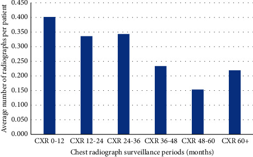
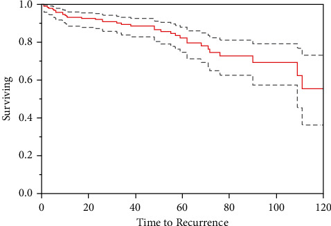
Results: 190 patients with ALT were included. Average age was 61.7 years and average follow-up was 58.6 months (24 to 180 months). MDM2 testing was positive in 88 patients (46.3%), and 102 (53.7%) did not receive MDM2 testing. 188 patients (98.9%) had marginal excision, and 127 (66.8%) had marginal or positive margins. Patients received an average of 0.9 CT scans and 1.3 chest radiographs over the surveillance period. 10-year metastasis-free survival was 100%, with no documented deaths from disease.
Conclusions: This study suggests that chest imaging does not have a significant role in PM surveillance following ALT excision, but advanced local imaging and chest surveillance may be considered in cases of local recurrence or concern for dedifferentiation.
SarcomaMedicine-Radiology, Nuclear Medicine and Imaging
CiteScore
5.00
自引率
0.00%
发文量
15
审稿时长
14 weeks
期刊介绍:
Sarcoma is dedicated to publishing papers covering all aspects of connective tissue oncology research. It brings together work from scientists and clinicians carrying out a broad range of research in this field, including the basic sciences, molecular biology and pathology and the clinical sciences of epidemiology, surgery, radiotherapy and chemotherapy. High-quality papers concerning the entire range of bone and soft tissue sarcomas in both adults and children, including Kaposi"s sarcoma, are published as well as preclinical and animal studies. This journal provides a central forum for the description of advances in diagnosis, assessment and treatment of this rarely seen, but often mismanaged, group of patients.


 求助内容:
求助内容: 应助结果提醒方式:


