Anatomical Variations of the Bifid Mandibular Canal on Panoramic Radiographs in Citizens from Zagreb, Croatia.
IF 1.4
Q3 DENTISTRY, ORAL SURGERY & MEDICINE
引用次数: 7
Abstract
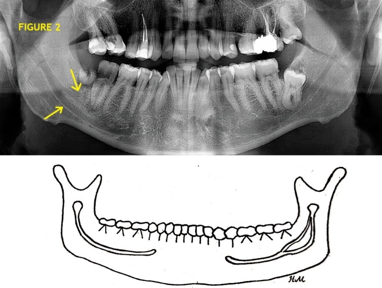
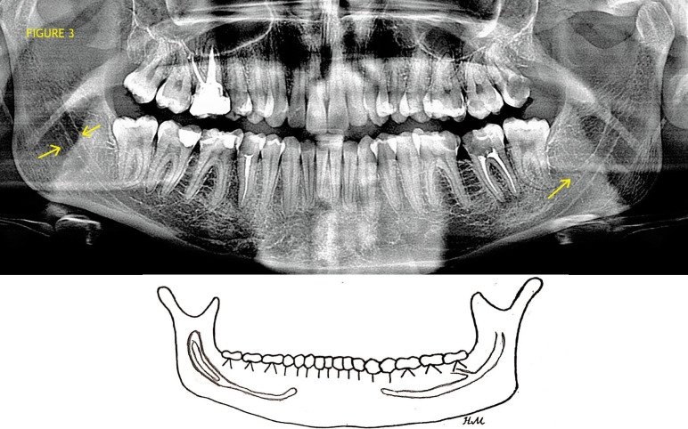
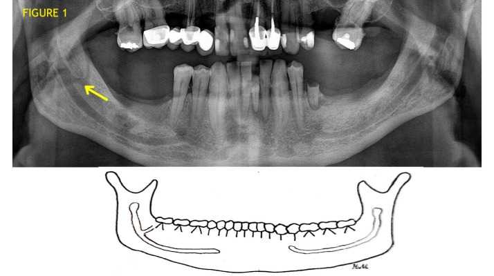
Background The bifid mandibular canal (BMC) is an anatomical variation with reported prevalence ranging from 0.08 to 65%. Identifying anatomical variations of mandibular canal is very important in order to prevent possible complications during oral surgical and other dental procedures. Objectives The aim of this study was to determine the prevalence and to classify the morphology of BMCs using digital panoramic radiographs. Material and methods A retrospective study was conducted that included 1008 digital panoramic radiographs (412 female and 596 male) used to identify the type of BMC. Panoramic radiographs were analyzed by three oral surgeons and one dentist, and BMCs were classified into six different types, 4 types according to Langlais et al. (types 1-4), and two new types (types 5 and 6) described by authors. Results The prevalence of BMC was 4.66% (n=47), with no significant differences in gender between BMC types (P=0.947; χ2=0.74). The prevalence of type 1 BMC was 0.79% (n=8), type 2 2.08% (n=21), type 3 0.30% (n=3), type 4 0% (n=0), type 5 0.89% (n=9) and type 6 0.60% (n=6). Conclusion This study revealed a relatively high prevalence of BMCs among Zagreb citizens. Furthermore, two new types of BMCs were described. These results stress the importance of a careful and thorough radiographic analysis prior to each invasive procedure in the mandible.
克罗地亚萨格勒布市民在全景x线片上的下颌双裂管解剖变异。
背景:下颌双裂管(BMC)是一种解剖变异,据报道发病率从0.08%到65%不等。识别下颌管的解剖变异是非常重要的,以防止可能的并发症在口腔外科和其他牙科手术。目的:本研究的目的是利用数字全景x线片确定bmc的患病率并对其形态进行分类。材料和方法:回顾性研究1008张用于识别BMC类型的数字全景x线片(女性412张,男性596张)。3名口腔外科医生和1名牙医对全景x线片进行分析,将bmc分为6种不同的类型,根据Langlais等人的4种类型(1-4型),以及作者描述的2种新类型(5型和6型)。结果:BMC患病率为4.66% (n=47), BMC类型间性别差异无统计学意义(P=0.947;χ2 = 0.74)。1型BMC患病率为0.79% (n=8), 2型为2.08% (n=21), 3型为0.30% (n=3), 4型为0% (n=0), 5型为0.89% (n=9), 6型为0.60% (n=6)。结论:本研究揭示了萨格勒布市民中bmc的患病率相对较高。此外,还描述了两种新的bmc类型。这些结果强调了在下颌骨每次侵入性手术之前进行仔细和彻底的放射学分析的重要性。
本文章由计算机程序翻译,如有差异,请以英文原文为准。
求助全文
约1分钟内获得全文
求助全文
来源期刊

Acta Stomatologica Croatica
DENTISTRY, ORAL SURGERY & MEDICINE-
CiteScore
2.40
自引率
28.60%
发文量
32
审稿时长
12 weeks
期刊介绍:
The Acta Stomatologica Croatica (ASCRO) is a leading scientific non-profit journal in the field of dental, oral and cranio-facial sciences during the past 44 years in Croatia. ASCRO publishes original scientific and clinical papers, preliminary communications, case reports, book reviews, letters to the editor and news. Review articles are published by invitation from the Editor-in-Chief by acclaimed professionals in distinct fields of dental medicine. All manuscripts are subjected to peer review process.
文献相关原料
| 公司名称 | 产品信息 | 采购帮参考价格 |
|---|
 求助内容:
求助内容: 应助结果提醒方式:


