{"title":"Structure of a retinal chromophore of dark-adapted middle rhodopsin as studied by solid-state nuclear magnetic resonance spectroscopy.","authors":"Izuru Kawamura, Hayato Seki, Seiya Tajima, Yoshiteru Makino, Arisu Shigeta, Takashi Okitsu, Akimori Wada, Akira Naito, Yuki Sudo","doi":"10.2142/biophysico.bppb-v18.019","DOIUrl":null,"url":null,"abstract":"<p><p>Middle rhodopsin (MR) found from the archaeon <i>Haloquadratum walsbyi</i> is evolutionarily located between two different types of rhodopsins, bacteriorhodopsin (BR) and sensory rhodopsin II (SRII). Some isomers of the chromophore retinal and the photochemical reaction of MR are markedly different from those of BR and SRII. In this study, to obtain the structural information regarding its active center (i.e., retinal), we subjected MR embedded in lipid bilayers to solid-state magic-angle spinning nuclear magnetic resonance (NMR) spectroscopy. The analysis of the isotropic <sup>13</sup>C chemical shifts of the retinal chromophore revealed the presence of three types of retinal configurations of dark-adapted MR: (13-<i>trans</i>, 15-<i>anti</i> (all-<i>trans</i>)), (13-<i>cis</i>, 15-<i>syn</i>), and 11-<i>cis</i> isomers. The higher field resonance of the 20-C methyl carbon in the all-<i>trans</i> retinal suggested that Trp182 in MR has an orientation that is different from that in other microbial rhodopsins, owing to the changes in steric hindrance associated with the 20-C methyl group in retinal. <sup>13</sup>Cζ signals of Tyr185 in MR for all-<i>trans</i> and 13-<i>cis</i>, 15-<i>syn</i> isomers were discretely observed, representing the difference in the hydrogen bond strength of Tyr185. Further, <sup>15</sup>N NMR analysis of the protonated Schiff base corresponding to the all-<i>trans</i> and 13-<i>cis</i>, 15-<i>syn</i> isomers in MR showed a strong electrostatic interaction with the counter ion. Therefore, the resulting structural information exhibited the property of stable retinal conformations of dark-adapted MR.</p>","PeriodicalId":8976,"journal":{"name":"Biophysics and Physicobiology","volume":" ","pages":"177-185"},"PeriodicalIF":0.0000,"publicationDate":"2021-07-14","publicationTypes":"Journal Article","fieldsOfStudy":null,"isOpenAccess":false,"openAccessPdf":"https://ftp.ncbi.nlm.nih.gov/pub/pmc/oa_pdf/81/90/18_177.PMC8354847.pdf","citationCount":"2","resultStr":null,"platform":"Semanticscholar","paperid":null,"PeriodicalName":"Biophysics and Physicobiology","FirstCategoryId":"1085","ListUrlMain":"https://doi.org/10.2142/biophysico.bppb-v18.019","RegionNum":0,"RegionCategory":null,"ArticlePicture":[],"TitleCN":null,"AbstractTextCN":null,"PMCID":null,"EPubDate":"2021/1/1 0:00:00","PubModel":"eCollection","JCR":"","JCRName":"","Score":null,"Total":0}
引用次数: 2
Abstract
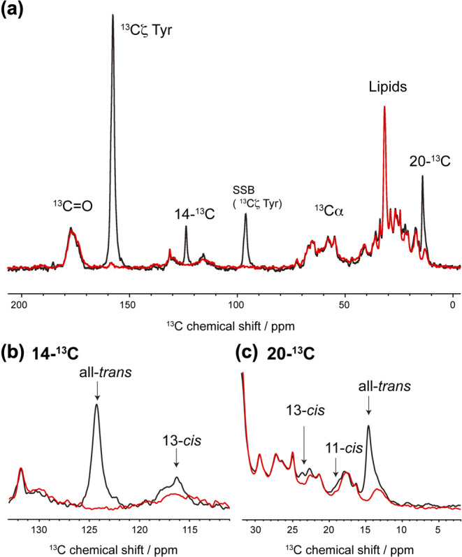
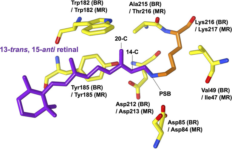
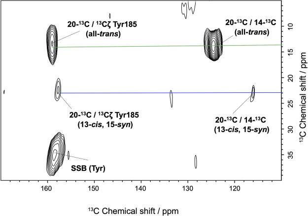
Middle rhodopsin (MR) found from the archaeon Haloquadratum walsbyi is evolutionarily located between two different types of rhodopsins, bacteriorhodopsin (BR) and sensory rhodopsin II (SRII). Some isomers of the chromophore retinal and the photochemical reaction of MR are markedly different from those of BR and SRII. In this study, to obtain the structural information regarding its active center (i.e., retinal), we subjected MR embedded in lipid bilayers to solid-state magic-angle spinning nuclear magnetic resonance (NMR) spectroscopy. The analysis of the isotropic 13C chemical shifts of the retinal chromophore revealed the presence of three types of retinal configurations of dark-adapted MR: (13-trans, 15-anti (all-trans)), (13-cis, 15-syn), and 11-cis isomers. The higher field resonance of the 20-C methyl carbon in the all-trans retinal suggested that Trp182 in MR has an orientation that is different from that in other microbial rhodopsins, owing to the changes in steric hindrance associated with the 20-C methyl group in retinal. 13Cζ signals of Tyr185 in MR for all-trans and 13-cis, 15-syn isomers were discretely observed, representing the difference in the hydrogen bond strength of Tyr185. Further, 15N NMR analysis of the protonated Schiff base corresponding to the all-trans and 13-cis, 15-syn isomers in MR showed a strong electrostatic interaction with the counter ion. Therefore, the resulting structural information exhibited the property of stable retinal conformations of dark-adapted MR.

 求助内容:
求助内容: 应助结果提醒方式:


