{"title":"Functional Imaging of the Cerebellum during Action Execution and Observation.","authors":"Vassilis Raos, Helen E Savaki","doi":"10.1093/texcom/tgab041","DOIUrl":null,"url":null,"abstract":"<p><p>We employed the <sup>14</sup>C-deoxyglucose autoradiographic method to map the activity in the cerebellar cortex of rhesus monkeys that performed forelimb movements either in the light or in the dark and of monkeys that observed forelimb movements executed by a human experimenter. The execution of forelimb movements, both in the light and in the dark, activated the forelimb representations in the cerebellar hemispheric extensions of 1) vermian lobules IV-VI and 2) vermian lobule VIIIB, ipsilaterally to the moving forelimb. Activations in the former forelimb representation involved both a paravermal and a lateral hemispheric region. Also, Crus II posterior in the ansiform lobule (the hemispheric expansion of lobule VIIB) was activated bilaterally by execution of movements in the light but not in the dark. Action observation activated the lateral-most region of the forelimb representation in the lateral hemispheric extension of vermian lobules IV-VI, as well as the crus II posterior, bilaterally. Our results demonstrate that the cerebellar cortex, in addition to its involvement in the generation of movement, is also recruited in the perception of observed movements. Moreover, our findings suggest a modularity gradient in the primate cerebellar cortex, which progresses from unimodal (medially) to multimodal (laterally) functional areas.</p>","PeriodicalId":72551,"journal":{"name":"Cerebral cortex communications","volume":"2 3","pages":"tgab041"},"PeriodicalIF":0.0000,"publicationDate":"2021-06-30","publicationTypes":"Journal Article","fieldsOfStudy":null,"isOpenAccess":false,"openAccessPdf":"https://www.ncbi.nlm.nih.gov/pmc/articles/PMC8366719/pdf/","citationCount":"2","resultStr":null,"platform":"Semanticscholar","paperid":null,"PeriodicalName":"Cerebral cortex communications","FirstCategoryId":"1085","ListUrlMain":"https://doi.org/10.1093/texcom/tgab041","RegionNum":0,"RegionCategory":null,"ArticlePicture":[],"TitleCN":null,"AbstractTextCN":null,"PMCID":null,"EPubDate":"2021/1/1 0:00:00","PubModel":"eCollection","JCR":"","JCRName":"","Score":null,"Total":0}
引用次数: 2
Abstract
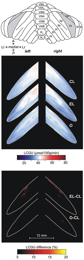
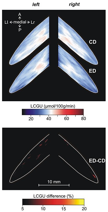
We employed the 14C-deoxyglucose autoradiographic method to map the activity in the cerebellar cortex of rhesus monkeys that performed forelimb movements either in the light or in the dark and of monkeys that observed forelimb movements executed by a human experimenter. The execution of forelimb movements, both in the light and in the dark, activated the forelimb representations in the cerebellar hemispheric extensions of 1) vermian lobules IV-VI and 2) vermian lobule VIIIB, ipsilaterally to the moving forelimb. Activations in the former forelimb representation involved both a paravermal and a lateral hemispheric region. Also, Crus II posterior in the ansiform lobule (the hemispheric expansion of lobule VIIB) was activated bilaterally by execution of movements in the light but not in the dark. Action observation activated the lateral-most region of the forelimb representation in the lateral hemispheric extension of vermian lobules IV-VI, as well as the crus II posterior, bilaterally. Our results demonstrate that the cerebellar cortex, in addition to its involvement in the generation of movement, is also recruited in the perception of observed movements. Moreover, our findings suggest a modularity gradient in the primate cerebellar cortex, which progresses from unimodal (medially) to multimodal (laterally) functional areas.

 求助内容:
求助内容: 应助结果提醒方式:


