Najma Latif, Ahmed Mahgoub, Mohamed Nagy, Padmini Sarathchandra, Magdi H Yacoub
{"title":"Severe degeneration of a sub-coronary pulmonary autograft in a young adult.","authors":"Najma Latif, Ahmed Mahgoub, Mohamed Nagy, Padmini Sarathchandra, Magdi H Yacoub","doi":"10.21542/gcsp.2021.14","DOIUrl":null,"url":null,"abstract":"<p><p><b>Background.</b> The pulmonary autograft is currently the best valve substitute in terms of longevity and performance. However, there is no agreement about the optimal method of insertion (sub-coronary position or freestanding root). <b>Objectives.</b> We sought to examine the clinical status, detailed imaging and morphometric changes in an explanted pulmonary autograft 22 years after sub-coronary implantation. <b>Methods.</b> A 30-year-old female underwent pulmonary autograft replacement of a severely stenotic valve at the age of 7 years, after presenting to us with signs of moderate to severe heart failure. She underwent clinical examination, detailed imaging including echocardiographic and CT examination with computerised image analysis. The explanted valve was examined by morphometry. <b>Results.</b> Clinical examination showed signs of heart failure (NYHA III). Trans-thoracic and trans-oesophageal 2D echo showed severe malfunction of both the aortic and pulmonary valves associated with dilatation and hypertrophy of both the right and left ventricles. Surgical correction was performed by replacing both the pulmonary and aortic valves with Medtronic 27mm Freestyle valves. The pulmonary autograft showed degeneration of the trilamellar layering of the leaflets, loss and disorganisation of GAGs, increased collagen with fibrotic overgrowth, and markers of fibrosis, inflammation, and calcification. Post-operative imaging showed good correction of the haemodynamic lesions. <b>Conclusion.</b> The pulmonary autograft implanted into the sub-coronary position presented with adverse remodelling, which was detrimental to the functionality and longevity of the valve. <b>Authorship.</b> NL, AM, MN all contributed equally to this paper.</p>","PeriodicalId":12669,"journal":{"name":"Global Cardiology Science & Practice","volume":"2021 2","pages":"e202114"},"PeriodicalIF":0.0000,"publicationDate":"2021-06-30","publicationTypes":"Journal Article","fieldsOfStudy":null,"isOpenAccess":false,"openAccessPdf":"https://www.ncbi.nlm.nih.gov/pmc/articles/PMC8272409/pdf/","citationCount":"0","resultStr":null,"platform":"Semanticscholar","paperid":null,"PeriodicalName":"Global Cardiology Science & Practice","FirstCategoryId":"1085","ListUrlMain":"https://doi.org/10.21542/gcsp.2021.14","RegionNum":0,"RegionCategory":null,"ArticlePicture":[],"TitleCN":null,"AbstractTextCN":null,"PMCID":null,"EPubDate":"","PubModel":"","JCR":"","JCRName":"","Score":null,"Total":0}
引用次数: 0
Abstract
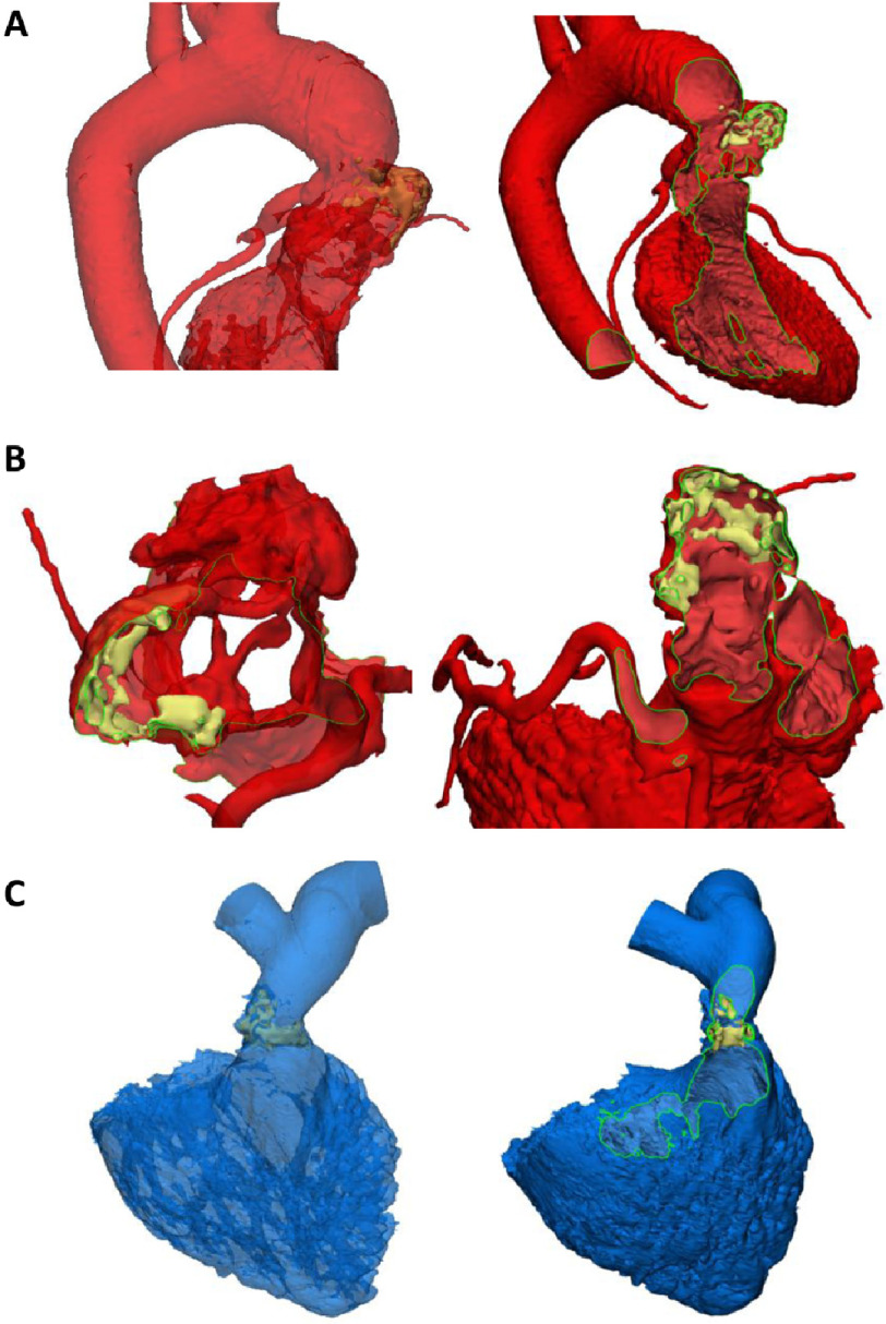
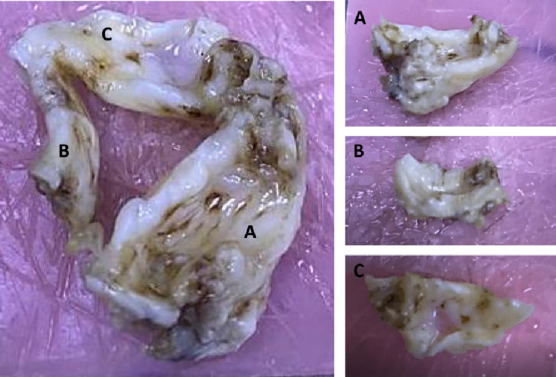
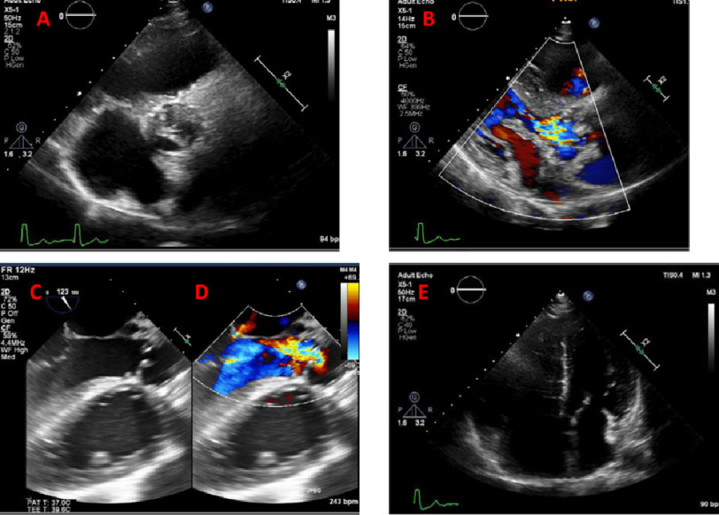
Background. The pulmonary autograft is currently the best valve substitute in terms of longevity and performance. However, there is no agreement about the optimal method of insertion (sub-coronary position or freestanding root). Objectives. We sought to examine the clinical status, detailed imaging and morphometric changes in an explanted pulmonary autograft 22 years after sub-coronary implantation. Methods. A 30-year-old female underwent pulmonary autograft replacement of a severely stenotic valve at the age of 7 years, after presenting to us with signs of moderate to severe heart failure. She underwent clinical examination, detailed imaging including echocardiographic and CT examination with computerised image analysis. The explanted valve was examined by morphometry. Results. Clinical examination showed signs of heart failure (NYHA III). Trans-thoracic and trans-oesophageal 2D echo showed severe malfunction of both the aortic and pulmonary valves associated with dilatation and hypertrophy of both the right and left ventricles. Surgical correction was performed by replacing both the pulmonary and aortic valves with Medtronic 27mm Freestyle valves. The pulmonary autograft showed degeneration of the trilamellar layering of the leaflets, loss and disorganisation of GAGs, increased collagen with fibrotic overgrowth, and markers of fibrosis, inflammation, and calcification. Post-operative imaging showed good correction of the haemodynamic lesions. Conclusion. The pulmonary autograft implanted into the sub-coronary position presented with adverse remodelling, which was detrimental to the functionality and longevity of the valve. Authorship. NL, AM, MN all contributed equally to this paper.

 求助内容:
求助内容: 应助结果提醒方式:


