A Levillain, S Ahmed, D-M Kaimaki, S Schuler, S Barros, D Labonte, J C Iatridis, N C Nowlan
{"title":"Prenatal muscle forces are necessary for vertebral segmentation and disc structure, but not for notochord involution in mice.","authors":"A Levillain, S Ahmed, D-M Kaimaki, S Schuler, S Barros, D Labonte, J C Iatridis, N C Nowlan","doi":"10.22203/eCM.v041a36","DOIUrl":null,"url":null,"abstract":"<p><p>Embryonic muscle forces are necessary for normal vertebral development and spinal curvature, but their involvement in intervertebral disc (IVD) development remains unclear. The aim of the current study was to determine how muscle contractions affect (1) notochord involution and vertebral segmentation, and (2) IVD development including the mechanical properties and morphology, as well as collagen fibre alignment in the annulus fibrosus. Muscular dysgenesis (mdg) mice were harvested at three prenatal stages: at Theiler Stage (TS)22 when notochord involution starts, at TS24 when involution is complete, and at TS27 when the IVD is formed. Vertebral and IVD development were characterised using histology, immunofluorescence, and indentation testing. The results revealed that notochord involution and vertebral segmentation occurred independently of muscle contractions between TS22 and TS24. However, in the absence of muscle contractions, we found vertebral fusion in the cervical region at TS27, along with (i) a displacement of the nucleus pulposus towards the dorsal side, (ii) a disruption of the structural arrangement of collagen in the annulus fibrosus, and (iii) an increase in viscous behaviour of the annulus fibrosus. These findings emphasise the important role of mechanical forces during IVD development, and demonstrate a critical role of muscle loading during development to enable proper annulus fibrosus formation. They further suggest a need for mechanical loading in the creation of fibre-reinforced tissue engineering replacement IVDs as a therapy for IVD degeneration.</p>","PeriodicalId":11849,"journal":{"name":"European cells & materials","volume":" ","pages":"558-575"},"PeriodicalIF":3.1000,"publicationDate":"2021-05-22","publicationTypes":"Journal Article","fieldsOfStudy":null,"isOpenAccess":false,"openAccessPdf":"https://ftp.ncbi.nlm.nih.gov/pub/pmc/oa_pdf/f6/6e/nihms-1718897.PMC8268087.pdf","citationCount":"0","resultStr":null,"platform":"Semanticscholar","paperid":null,"PeriodicalName":"European cells & materials","FirstCategoryId":"5","ListUrlMain":"https://doi.org/10.22203/eCM.v041a36","RegionNum":3,"RegionCategory":"医学","ArticlePicture":[],"TitleCN":null,"AbstractTextCN":null,"PMCID":null,"EPubDate":"","PubModel":"","JCR":"Q3","JCRName":"CELL & TISSUE ENGINEERING","Score":null,"Total":0}
引用次数: 0
Abstract
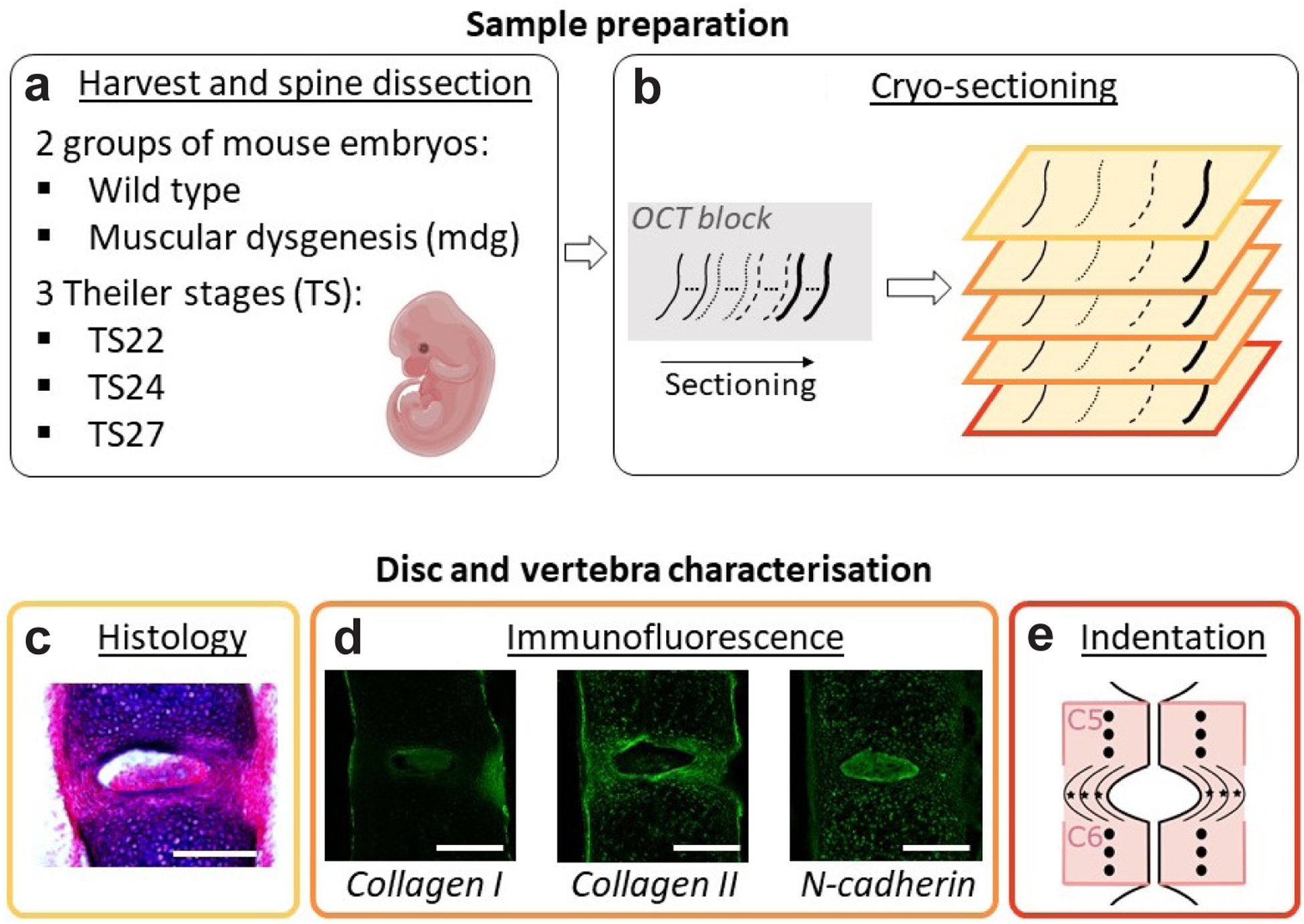
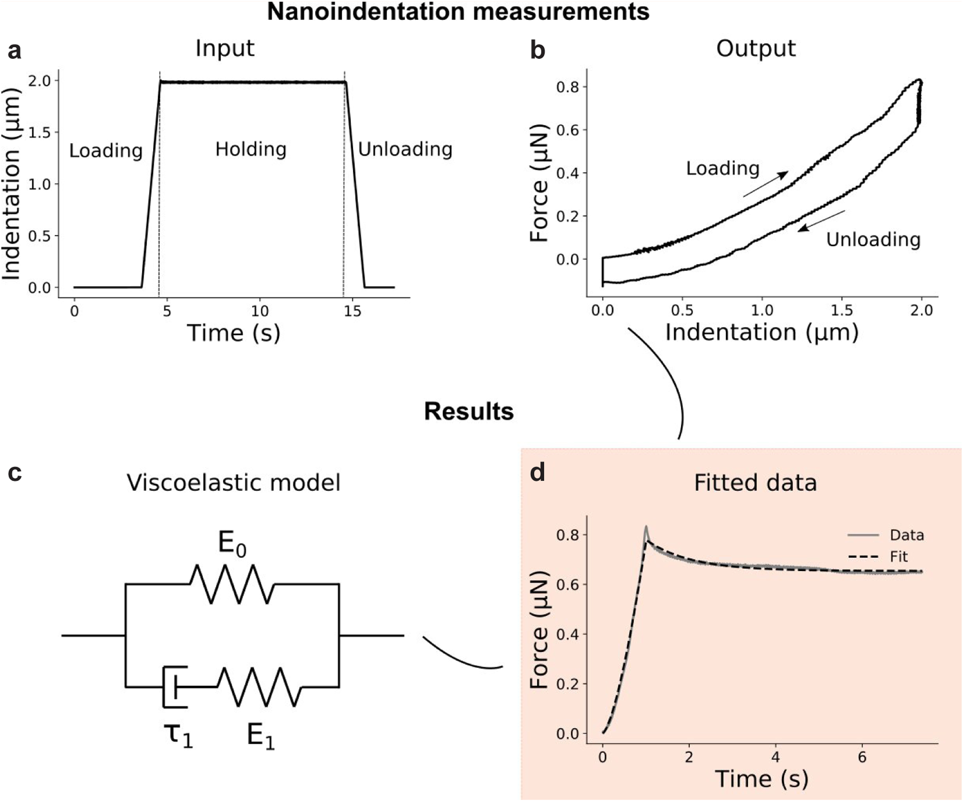
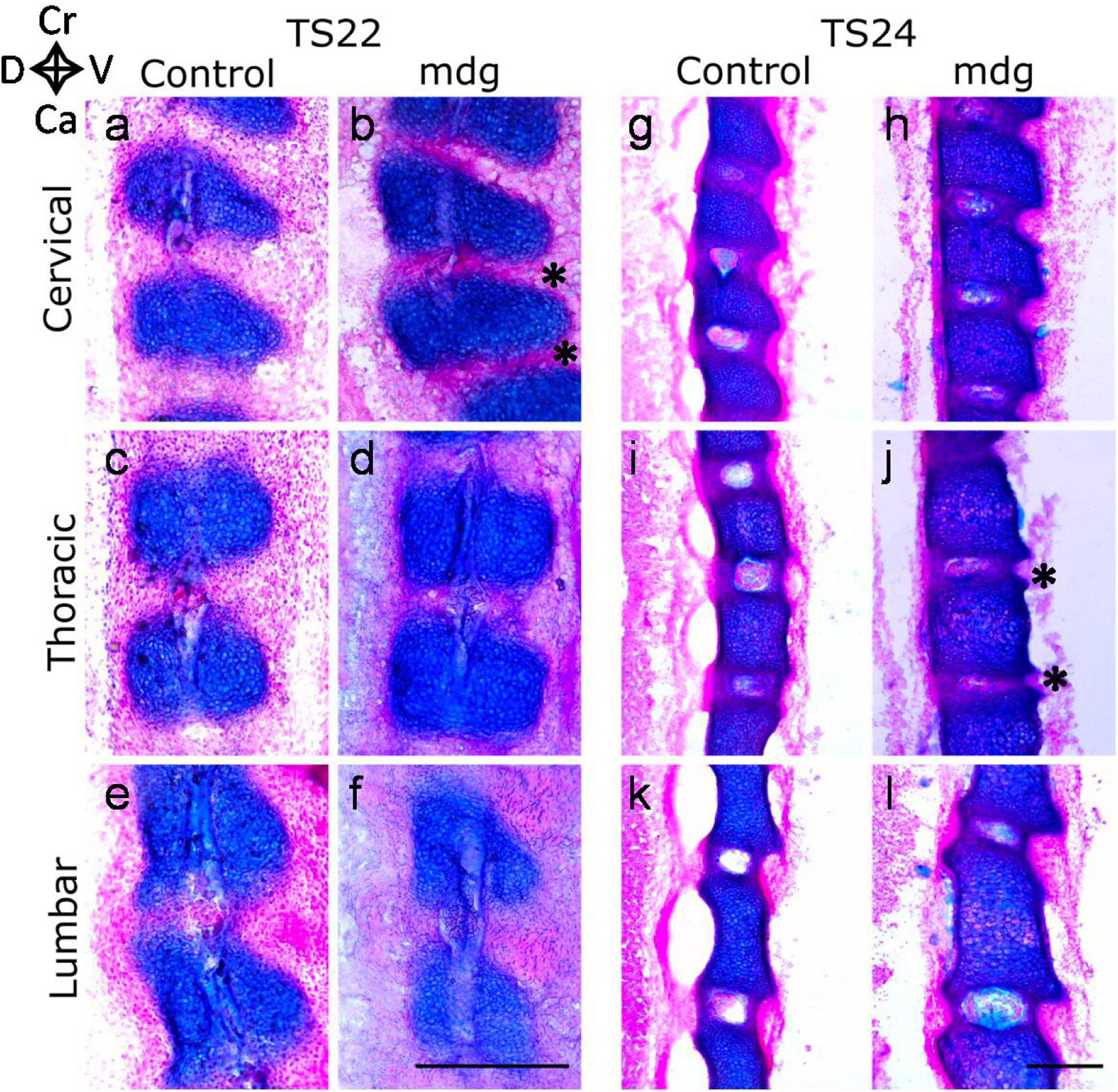
Embryonic muscle forces are necessary for normal vertebral development and spinal curvature, but their involvement in intervertebral disc (IVD) development remains unclear. The aim of the current study was to determine how muscle contractions affect (1) notochord involution and vertebral segmentation, and (2) IVD development including the mechanical properties and morphology, as well as collagen fibre alignment in the annulus fibrosus. Muscular dysgenesis (mdg) mice were harvested at three prenatal stages: at Theiler Stage (TS)22 when notochord involution starts, at TS24 when involution is complete, and at TS27 when the IVD is formed. Vertebral and IVD development were characterised using histology, immunofluorescence, and indentation testing. The results revealed that notochord involution and vertebral segmentation occurred independently of muscle contractions between TS22 and TS24. However, in the absence of muscle contractions, we found vertebral fusion in the cervical region at TS27, along with (i) a displacement of the nucleus pulposus towards the dorsal side, (ii) a disruption of the structural arrangement of collagen in the annulus fibrosus, and (iii) an increase in viscous behaviour of the annulus fibrosus. These findings emphasise the important role of mechanical forces during IVD development, and demonstrate a critical role of muscle loading during development to enable proper annulus fibrosus formation. They further suggest a need for mechanical loading in the creation of fibre-reinforced tissue engineering replacement IVDs as a therapy for IVD degeneration.
期刊介绍:
eCM provides an interdisciplinary forum for publication of preclinical research in the musculoskeletal field (Trauma, Maxillofacial (including dental), Spine and Orthopaedics).
The clinical relevance of the work must be briefly mentioned within the abstract, and in more detail in the paper. Poor abstracts which do not concisely cover the paper contents will not be sent for review. Incremental steps in research will not be entertained by eCM journal.Cross-disciplinary papers that go across our scope areas are welcomed.

 求助内容:
求助内容: 应助结果提醒方式:


