Pasquale Loiudice, Marco Pellegrini, Michele Marinò, Barbara Mazzi, Ilaria Ionni, Giuseppe Covello, Michele Figus, Marco Nardi, Giamberto Casini
{"title":"Choroidal vascularity index in thyroid-associated ophthalmopathy: a cross-sectional study.","authors":"Pasquale Loiudice, Marco Pellegrini, Michele Marinò, Barbara Mazzi, Ilaria Ionni, Giuseppe Covello, Michele Figus, Marco Nardi, Giamberto Casini","doi":"10.1186/s40662-021-00242-6","DOIUrl":null,"url":null,"abstract":"<p><strong>Background: </strong>Hemodynamic changes have been observed in patients with Graves' disease. The aim of our study was to evaluate choroidal vascular change using the choroidal vascularity index (CVI) in patients with thyroid-associated ophthalmopathy (TAO).</p><p><strong>Methods: </strong>In this cross-sectional observational study, 40 patients affected by TAO were recruited. Forty healthy individuals, matched for age and sex, served as controls. Foveal enhanced-depth imaging optical coherence tomography scans were obtained from all participants. Images were binarized using the ImageJ software and luminal area (LA) and total choroidal area (TCA) were measured. CVI was calculated as the proportion of LA to TCA. The relation between CVI or subfoveal choroidal thickness (SFCT) and clinical activity score, exophthalmometric value, diplopia status, gender, and age was evaluated.</p><p><strong>Results: </strong>CVI was significantly higher in patients with TAO (P = 0.004). No significant difference was observed in SFCT (P = 0.200) and TCA (P = 0.153) comparing TAO patients and healthy controls. LA was significantly higher in TAO group (P = 0.045). On multiple regression analysis, CVI was associated with TCA (P = 0.043). No association was found between SFCT or CVI and TCA, clinical activity score, exophthalmometric value, Inami value, diplopia status, gender or age (P > 0.05).</p><p><strong>Conclusions: </strong>This is the first study that has demonstrated an increase in CVI in eyes with TAO compared with healthy controls and has assessed its association with clinical features.</p>","PeriodicalId":520624,"journal":{"name":"Eye and vision (London, England)","volume":" ","pages":"18"},"PeriodicalIF":0.0000,"publicationDate":"2021-04-30","publicationTypes":"Journal Article","fieldsOfStudy":null,"isOpenAccess":false,"openAccessPdf":"https://sci-hub-pdf.com/10.1186/s40662-021-00242-6","citationCount":"12","resultStr":null,"platform":"Semanticscholar","paperid":null,"PeriodicalName":"Eye and vision (London, England)","FirstCategoryId":"3","ListUrlMain":"https://doi.org/10.1186/s40662-021-00242-6","RegionNum":0,"RegionCategory":null,"ArticlePicture":[],"TitleCN":null,"AbstractTextCN":null,"PMCID":null,"EPubDate":"","PubModel":"","JCR":"","JCRName":"","Score":null,"Total":0}
引用次数: 12
Abstract
Background: Hemodynamic changes have been observed in patients with Graves' disease. The aim of our study was to evaluate choroidal vascular change using the choroidal vascularity index (CVI) in patients with thyroid-associated ophthalmopathy (TAO).
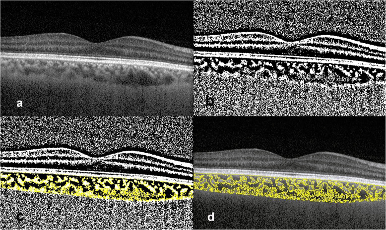
Methods: In this cross-sectional observational study, 40 patients affected by TAO were recruited. Forty healthy individuals, matched for age and sex, served as controls. Foveal enhanced-depth imaging optical coherence tomography scans were obtained from all participants. Images were binarized using the ImageJ software and luminal area (LA) and total choroidal area (TCA) were measured. CVI was calculated as the proportion of LA to TCA. The relation between CVI or subfoveal choroidal thickness (SFCT) and clinical activity score, exophthalmometric value, diplopia status, gender, and age was evaluated.
Results: CVI was significantly higher in patients with TAO (P = 0.004). No significant difference was observed in SFCT (P = 0.200) and TCA (P = 0.153) comparing TAO patients and healthy controls. LA was significantly higher in TAO group (P = 0.045). On multiple regression analysis, CVI was associated with TCA (P = 0.043). No association was found between SFCT or CVI and TCA, clinical activity score, exophthalmometric value, Inami value, diplopia status, gender or age (P > 0.05).
Conclusions: This is the first study that has demonstrated an increase in CVI in eyes with TAO compared with healthy controls and has assessed its association with clinical features.

 求助内容:
求助内容: 应助结果提醒方式:


