Yunhai Tu, Mingna Xu, Andy D Kim, Michael T M Wang, Zhaoqi Pan, Wencan Wu
{"title":"Modified endoscopic transnasal orbital apex decompression in dysthyroid optic neuropathy.","authors":"Yunhai Tu, Mingna Xu, Andy D Kim, Michael T M Wang, Zhaoqi Pan, Wencan Wu","doi":"10.1186/s40662-021-00238-2","DOIUrl":null,"url":null,"abstract":"<p><strong>Background: </strong>To describe the surgical technique and assess the clinical efficacy and safety of modified endoscopic transnasal orbital apex decompression in the treatment of dysthyroid optic neuropathy.</p><p><strong>Methods: </strong>In this retrospective research, forty-two subjects (74 orbits) who underwent modified endoscopic transnasal orbital apex decompression for the treatment of dysthyroid optic neuropathy were enrolled. Preoperative and postoperative best-corrected visual acuity (BCVA), visual field mean deviation (MD), Hertel exophthalmometry, and new onset diplopia were assessed before and after the intervention. The Wilcoxon test was used for differential analysis. Linear mixed-models' analyses were conducted to assess the potential predictors for BCVA change.</p><p><strong>Results: </strong>Postoperatively, the mean BCVA improved from 0.70 ± 0.62 logMAR to 0.22 ± 0.33 logMAR. BCVA significantly improved in 69 eyes (93%), remained stable in 4 eyes (5%) and deteriorated in 1 eye (1%). MD of visual fields improved from -13.73 ± 9.22 dB to -7.23 ± 7.04 dB. Proptosis decreased from 19.57 ± 3.38 mm to 16.35 ± 3.01 mm. Preoperative BCVA, MD of visual fields and medical rectus diameter were independent factors associated with improvements in BCVA (P < 0.05) by linear mixed-models' analyses. Eighteen patients (42.9%) developed new diplopia postoperatively.</p><p><strong>Conclusion: </strong>Modified endoscopic transnasal orbital apex decompression effectively restores vision in dysthyroid optic neuropathy.</p>","PeriodicalId":520624,"journal":{"name":"Eye and vision (London, England)","volume":" ","pages":"19"},"PeriodicalIF":0.0000,"publicationDate":"2021-04-28","publicationTypes":"Journal Article","fieldsOfStudy":null,"isOpenAccess":false,"openAccessPdf":"https://sci-hub-pdf.com/10.1186/s40662-021-00238-2","citationCount":"6","resultStr":null,"platform":"Semanticscholar","paperid":null,"PeriodicalName":"Eye and vision (London, England)","FirstCategoryId":"3","ListUrlMain":"https://doi.org/10.1186/s40662-021-00238-2","RegionNum":0,"RegionCategory":null,"ArticlePicture":[],"TitleCN":null,"AbstractTextCN":null,"PMCID":null,"EPubDate":"","PubModel":"","JCR":"","JCRName":"","Score":null,"Total":0}
引用次数: 6
Abstract
Background: To describe the surgical technique and assess the clinical efficacy and safety of modified endoscopic transnasal orbital apex decompression in the treatment of dysthyroid optic neuropathy.
Methods: In this retrospective research, forty-two subjects (74 orbits) who underwent modified endoscopic transnasal orbital apex decompression for the treatment of dysthyroid optic neuropathy were enrolled. Preoperative and postoperative best-corrected visual acuity (BCVA), visual field mean deviation (MD), Hertel exophthalmometry, and new onset diplopia were assessed before and after the intervention. The Wilcoxon test was used for differential analysis. Linear mixed-models' analyses were conducted to assess the potential predictors for BCVA change.
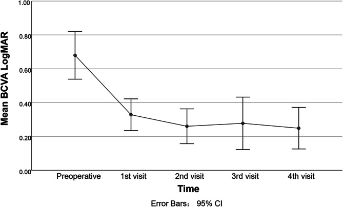
Results: Postoperatively, the mean BCVA improved from 0.70 ± 0.62 logMAR to 0.22 ± 0.33 logMAR. BCVA significantly improved in 69 eyes (93%), remained stable in 4 eyes (5%) and deteriorated in 1 eye (1%). MD of visual fields improved from -13.73 ± 9.22 dB to -7.23 ± 7.04 dB. Proptosis decreased from 19.57 ± 3.38 mm to 16.35 ± 3.01 mm. Preoperative BCVA, MD of visual fields and medical rectus diameter were independent factors associated with improvements in BCVA (P < 0.05) by linear mixed-models' analyses. Eighteen patients (42.9%) developed new diplopia postoperatively.
Conclusion: Modified endoscopic transnasal orbital apex decompression effectively restores vision in dysthyroid optic neuropathy.

 求助内容:
求助内容: 应助结果提醒方式:


