{"title":"Primary intracranial leiomyosarcoma presenting with frontal bone mass: a case report.","authors":"Shaghayegh Kamian, Abdolali Ebrahimi, Kaveh Ebrahim Zadeh, Behnaz Behzadi","doi":"10.3857/roj.2020.00577","DOIUrl":null,"url":null,"abstract":"<p><p>Primary intracranial mesenchymal neoplasms are rare tumors. These tumors are usually metastatic disease from other primary sites. We presented a 31-year-old man with a 6-month history of gradually enlarging frontal mass and positional headache. There was no other symptom demonstrating other organs' involvement. The patient underwent an uncomplicated craniotomy with clear surgical margins. The pathology review and the immunohistochemistry staining confirmed leiomyosarcoma grade II. We prescribed radiation therapy with tumor dose of 60 Gy in 30 fractions with conformal treatment planning to the tumor bed. As this disease has a high potency for metastasis, we advised four courses of single agent doxorubicin chemotherapy 75 mg/m2 every 4 weeks starting one month after the end of radiotherapy. In the last follow-up visit 34 months later, the patient was disease free in physical exam and imaging findings.</p>","PeriodicalId":46572,"journal":{"name":"Radiation Oncology Journal","volume":"38 4","pages":"282-286"},"PeriodicalIF":1.8000,"publicationDate":"2020-12-01","publicationTypes":"Journal Article","fieldsOfStudy":null,"isOpenAccess":false,"openAccessPdf":"https://ftp.ncbi.nlm.nih.gov/pub/pmc/oa_pdf/25/af/roj-2020-00577.PMC7785836.pdf","citationCount":"2","resultStr":null,"platform":"Semanticscholar","paperid":null,"PeriodicalName":"Radiation Oncology Journal","FirstCategoryId":"1085","ListUrlMain":"https://doi.org/10.3857/roj.2020.00577","RegionNum":0,"RegionCategory":null,"ArticlePicture":[],"TitleCN":null,"AbstractTextCN":null,"PMCID":null,"EPubDate":"2020/12/2 0:00:00","PubModel":"Epub","JCR":"Q3","JCRName":"ONCOLOGY","Score":null,"Total":0}
引用次数: 2
Abstract
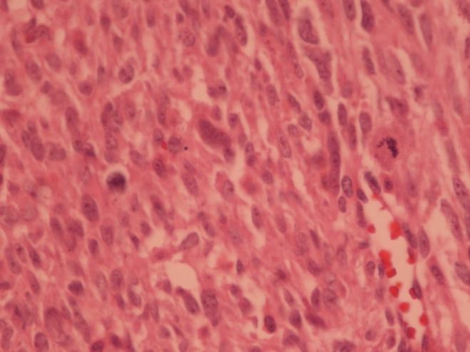
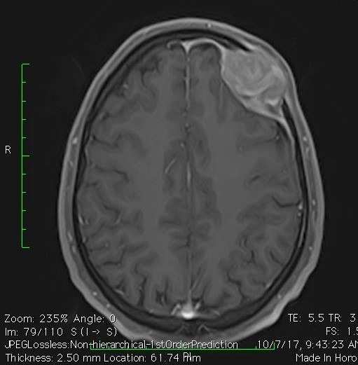
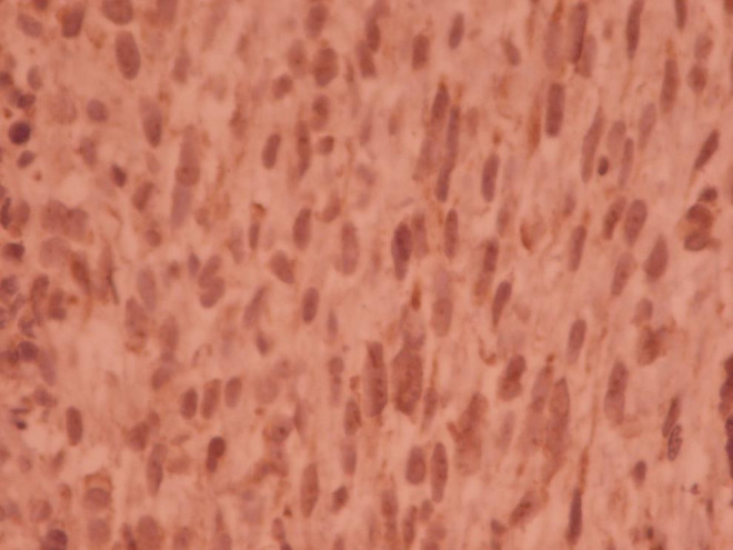
Primary intracranial mesenchymal neoplasms are rare tumors. These tumors are usually metastatic disease from other primary sites. We presented a 31-year-old man with a 6-month history of gradually enlarging frontal mass and positional headache. There was no other symptom demonstrating other organs' involvement. The patient underwent an uncomplicated craniotomy with clear surgical margins. The pathology review and the immunohistochemistry staining confirmed leiomyosarcoma grade II. We prescribed radiation therapy with tumor dose of 60 Gy in 30 fractions with conformal treatment planning to the tumor bed. As this disease has a high potency for metastasis, we advised four courses of single agent doxorubicin chemotherapy 75 mg/m2 every 4 weeks starting one month after the end of radiotherapy. In the last follow-up visit 34 months later, the patient was disease free in physical exam and imaging findings.

 求助内容:
求助内容: 应助结果提醒方式:


