{"title":"Is it possible to predict the side of hepatic metastases according to the primary location of colorectal cancer?","authors":"Ayşegül Gürsoy Çoruh, Çağlar Uzun, Esra Bozca, Büşra Bozca, İhsan Batuhan Demir, Hilal Gülseren Atasever, Berna Göçtürk, Batuhan Bakırarar, Cihangir Akyol","doi":"10.5114/pjr.2020.99848","DOIUrl":null,"url":null,"abstract":"<p><strong>Purpose: </strong>The aim of this study was to investigate the impact of the primary location of colorectal adenocarcinoma on the lobar distribution of its hepatic metastases based on the streamline hypothesis.</p><p><strong>Material and methods: </strong>The hospital database was utilised to identify the colorectal cancer patients. Eighty-six patients diagnosed with colorectal adenocarcinoma, who had hepatic metastases on the initial diagnostic stage or on the follow-up investigations, were enrolled the study. Computed tomography (CT) images of the study population were reviewed for the primary location of the colorectal tumour, and the side and number of hepatic metastases.</p><p><strong>Results: </strong>A total of 481 metastases were counted on CT from 22 right-sided and 64 left-sided colon tumours. The ratio of right-to-left hemiliver involvement was 1.97 : 1 for whole study population. The right-to-left ratio was calculated as 1.55 : 1 for right colon tumours and 2.17 : 1 for left colon tumours (<i>p</i> = 0.106). In the subgroup analysis with unilobar metastatic patients, again there was no significant difference in terms of the colorectal tumours' primary location (<i>p</i> = 0.325).</p><p><strong>Conclusions: </strong>The lobar distribution of hepatic metastases from colorectal adenocarcinoma may not be associated with the primary tumour localisation.</p>","PeriodicalId":47128,"journal":{"name":"Polish Journal of Radiology","volume":"85 ","pages":"e595-e599"},"PeriodicalIF":0.9000,"publicationDate":"2020-10-27","publicationTypes":"Journal Article","fieldsOfStudy":null,"isOpenAccess":false,"openAccessPdf":"https://ftp.ncbi.nlm.nih.gov/pub/pmc/oa_pdf/da/6d/PJR-85-42037.PMC7654317.pdf","citationCount":"0","resultStr":null,"platform":"Semanticscholar","paperid":null,"PeriodicalName":"Polish Journal of Radiology","FirstCategoryId":"1085","ListUrlMain":"https://doi.org/10.5114/pjr.2020.99848","RegionNum":0,"RegionCategory":null,"ArticlePicture":[],"TitleCN":null,"AbstractTextCN":null,"PMCID":null,"EPubDate":"2020/1/1 0:00:00","PubModel":"eCollection","JCR":"Q4","JCRName":"RADIOLOGY, NUCLEAR MEDICINE & MEDICAL IMAGING","Score":null,"Total":0}
引用次数: 0
Abstract
Purpose: The aim of this study was to investigate the impact of the primary location of colorectal adenocarcinoma on the lobar distribution of its hepatic metastases based on the streamline hypothesis.
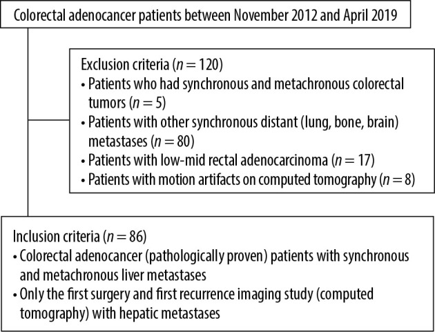
Material and methods: The hospital database was utilised to identify the colorectal cancer patients. Eighty-six patients diagnosed with colorectal adenocarcinoma, who had hepatic metastases on the initial diagnostic stage or on the follow-up investigations, were enrolled the study. Computed tomography (CT) images of the study population were reviewed for the primary location of the colorectal tumour, and the side and number of hepatic metastases.
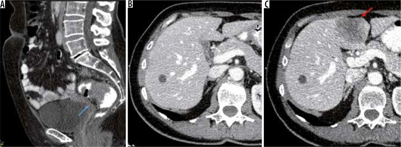
Results: A total of 481 metastases were counted on CT from 22 right-sided and 64 left-sided colon tumours. The ratio of right-to-left hemiliver involvement was 1.97 : 1 for whole study population. The right-to-left ratio was calculated as 1.55 : 1 for right colon tumours and 2.17 : 1 for left colon tumours (p = 0.106). In the subgroup analysis with unilobar metastatic patients, again there was no significant difference in terms of the colorectal tumours' primary location (p = 0.325).
Conclusions: The lobar distribution of hepatic metastases from colorectal adenocarcinoma may not be associated with the primary tumour localisation.

 求助内容:
求助内容: 应助结果提醒方式:


