Stephen W Sawyer, Kairui Zhang, Jason A Horton, Pranav Soman
{"title":"Perfusion-based co-culture model system for bone tissue engineering.","authors":"Stephen W Sawyer, Kairui Zhang, Jason A Horton, Pranav Soman","doi":"10.3934/bioeng.2020009","DOIUrl":null,"url":null,"abstract":"<p><p>In this work, we report on a perfusion-based co-culture system that could be used for bone tissue engineering applications. The model system is created using a combination of Primary Human Umbilical Vein Endothelial Cells (HUVECs) and osteoblast-like Saos-2 cells encapsulated within a Gelatin Methacrylate (GelMA)-collagen hydrogel blend contained within 3D printed, perfusable constructs. The constructs contain dual channels, within a custom-built bioreactor, that were perfused with osteogenic media for up to two weeks in order to induce mineral deposition. Mineral deposition in constructs containing only HUVECs, only Saos-2 cells, or a combination thereof was quantified by microCT to determine if the combination of endothelial cells and bone-like cells increased mineral deposition. Histological and fluorescent staining was used to verify mineral deposition and cellular function both along and between the perfused channels. While there was not a quantifiable difference in the amount of mineral deposited in Saos-2 only versus Saos-2 plus HUVEC samples, the location of the deposited mineral differed dramatically between the groups and indicated that the addition of HUVECs within the GelMA matrix allowed Saos-2 cells, in diffusion limited regions of the construct, to deposit bone mineral. This work serves as a model on how to create perfusable bone tissue engineering constructs using a combination of 3D printing and cellular co-cultures.</p>","PeriodicalId":45029,"journal":{"name":"AIMS Bioengineering","volume":"7 2","pages":"91-105"},"PeriodicalIF":1.2000,"publicationDate":"2020-01-01","publicationTypes":"Journal Article","fieldsOfStudy":null,"isOpenAccess":false,"openAccessPdf":"https://www.ncbi.nlm.nih.gov/pmc/articles/PMC7643915/pdf/","citationCount":"6","resultStr":null,"platform":"Semanticscholar","paperid":null,"PeriodicalName":"AIMS Bioengineering","FirstCategoryId":"1085","ListUrlMain":"https://doi.org/10.3934/bioeng.2020009","RegionNum":0,"RegionCategory":null,"ArticlePicture":[],"TitleCN":null,"AbstractTextCN":null,"PMCID":null,"EPubDate":"2020/5/29 0:00:00","PubModel":"Epub","JCR":"Q4","JCRName":"ENGINEERING, BIOMEDICAL","Score":null,"Total":0}
引用次数: 6
Abstract
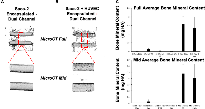
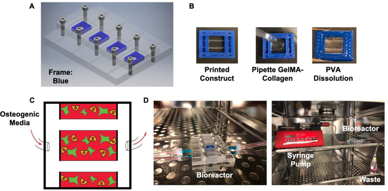
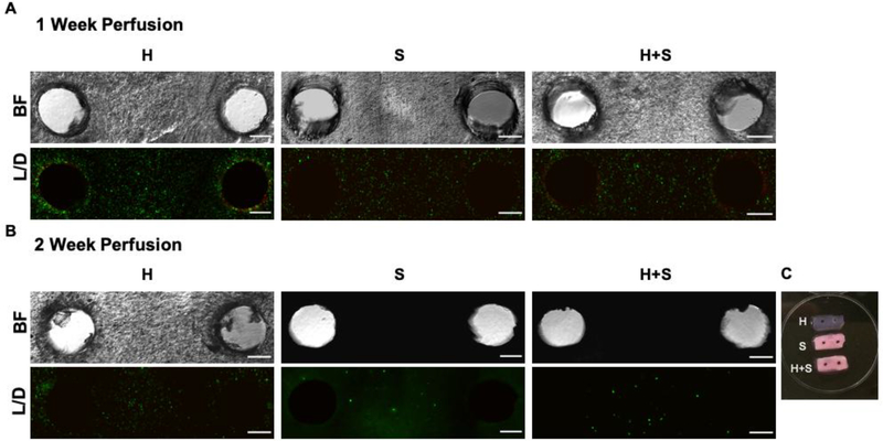
In this work, we report on a perfusion-based co-culture system that could be used for bone tissue engineering applications. The model system is created using a combination of Primary Human Umbilical Vein Endothelial Cells (HUVECs) and osteoblast-like Saos-2 cells encapsulated within a Gelatin Methacrylate (GelMA)-collagen hydrogel blend contained within 3D printed, perfusable constructs. The constructs contain dual channels, within a custom-built bioreactor, that were perfused with osteogenic media for up to two weeks in order to induce mineral deposition. Mineral deposition in constructs containing only HUVECs, only Saos-2 cells, or a combination thereof was quantified by microCT to determine if the combination of endothelial cells and bone-like cells increased mineral deposition. Histological and fluorescent staining was used to verify mineral deposition and cellular function both along and between the perfused channels. While there was not a quantifiable difference in the amount of mineral deposited in Saos-2 only versus Saos-2 plus HUVEC samples, the location of the deposited mineral differed dramatically between the groups and indicated that the addition of HUVECs within the GelMA matrix allowed Saos-2 cells, in diffusion limited regions of the construct, to deposit bone mineral. This work serves as a model on how to create perfusable bone tissue engineering constructs using a combination of 3D printing and cellular co-cultures.

 求助内容:
求助内容: 应助结果提醒方式:


