Cor triatriatrum or divided left atrium presenting as mitral stenosis in an adult patient.
IF 2.4
Q2 CARDIAC & CARDIOVASCULAR SYSTEMS
引用次数: 0
Abstract
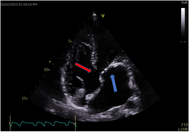
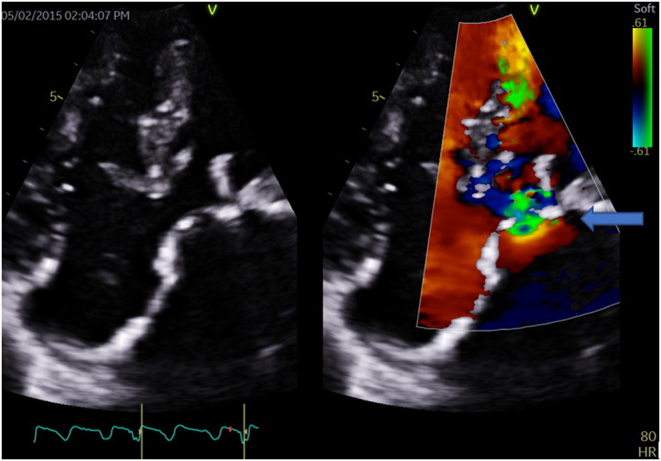
A 26-year-old male patient presented to the hospital with a 2-month history of progressive dyspnoea. He denied chest pain, coughing, orthopnoea, paroxysmal nocturnal dyspnoea, syncope or pre-syncope. He had no other significant comorbidities and he was not on any chronic medication. His cardiovascular examination revealed an undisplaced apex beat with a parasternal heave and a loud second heart sound (P2) suggestive of pulmonary hypertension. On auscultation, the first heart sound was normal with a loud second heart sound and a diastolic rumble. Given these findings, a clinical diagnosis of severe mitral stenosis with pulmonary hypertension was made. ECG was atypical for mitral stenosis, it revealed a dilated LA with left axis deviation due to a left anterior hemiblock. No features of right ventricular hypertrophy was noted. To our surprise, echocardiographic evaluation (Figs 1 and 2) revealed a primum ASD with the normal function of the left and right atrioventricular (AV) valve. A left-sided supra-valvular ridge or divided left atrium was identified with peak and mean gradients of 43/21 mmHg, respectively (Fig. 3). Video 1 is an apical four-chamber view of the defect, pre-operatively. -20-0016 ID: 20-0016


成人三房心或左心房分裂表现为二尖瓣狭窄。
本文章由计算机程序翻译,如有差异,请以英文原文为准。
求助全文
约1分钟内获得全文
求助全文
来源期刊

Echo Research and Practice
CARDIAC & CARDIOVASCULAR SYSTEMS-
CiteScore
6.70
自引率
12.70%
发文量
11
审稿时长
8 weeks
期刊介绍:
Echo Research and Practice aims to be the premier international journal for physicians, sonographers, nurses and other allied health professionals practising echocardiography and other cardiac imaging modalities. This open-access journal publishes quality clinical and basic research, reviews, videos, education materials and selected high-interest case reports and videos across all echocardiography modalities and disciplines, including paediatrics, anaesthetics, general practice, acute medicine and intensive care. Multi-modality studies primarily featuring the use of cardiac ultrasound in clinical practice, in association with Cardiac Computed Tomography, Cardiovascular Magnetic Resonance or Nuclear Cardiology are of interest. Topics include, but are not limited to: 2D echocardiography 3D echocardiography Comparative imaging techniques – CCT, CMR and Nuclear Cardiology Congenital heart disease, including foetal echocardiography Contrast echocardiography Critical care echocardiography Deformation imaging Doppler echocardiography Interventional echocardiography Intracardiac echocardiography Intraoperative echocardiography Prosthetic valves Stress echocardiography Technical innovations Transoesophageal echocardiography Valve disease.
 求助内容:
求助内容: 应助结果提醒方式:


