Paravalvular leak closure with real time transesophageal echocardiography and fluoroscopy fusion.
IF 1.5
Q3 CARDIAC & CARDIOVASCULAR SYSTEMS
JRSM Cardiovascular Disease
Pub Date : 2020-09-13
eCollection Date: 2020-01-01
DOI:10.1177/2048004020947290
引用次数: 5
Abstract
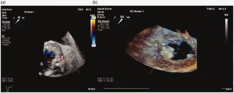
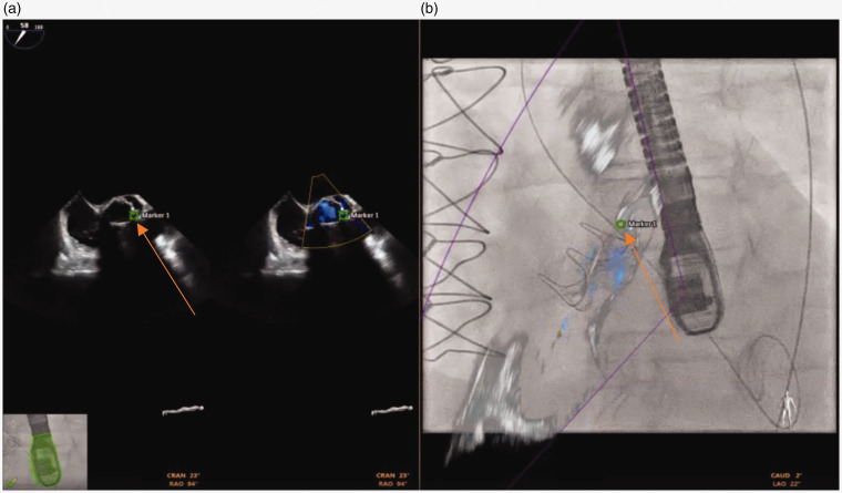
Transcatheter technology has been increasingly used for paravalvular leak closure. We report the use of "Fusion Technology" (EchoNaviagator, Phillips, Tustin, CA) that combines real-time 2 and 3 dimensional trans-esophageal echocardiography with fluoroscopy imaging to facilitate paravalvular leak closure. This could help to identify the exact site, size, depth and shape of the paravalvular leak for proper positioning of the occluder device, which may result in saving time and effort.
实时经食管超声心动图与透视融合治疗瓣旁漏。
经导管技术越来越多地用于瓣旁泄漏闭合。我们报告使用“融合技术”(EchoNaviagator, Phillips, Tustin, CA),将实时二维和三维经食管超声心动图与透视成像相结合,以促进瓣旁泄漏闭合。这有助于确定瓣旁泄漏的确切位置、大小、深度和形状,以便正确定位封堵器装置,从而节省时间和精力。
本文章由计算机程序翻译,如有差异,请以英文原文为准。
求助全文
约1分钟内获得全文
求助全文

 求助内容:
求助内容: 应助结果提醒方式:


