Taher Eleiwa, Amr Elsawy, Eyüp Özcan, Mohamed Abou Shousha
{"title":"Automated diagnosis and staging of Fuchs' endothelial cell corneal dystrophy using deep learning.","authors":"Taher Eleiwa, Amr Elsawy, Eyüp Özcan, Mohamed Abou Shousha","doi":"10.1186/s40662-020-00209-z","DOIUrl":null,"url":null,"abstract":"<p><strong>Background: </strong>To describe the diagnostic performance of a deep learning algorithm in discriminating early-stage Fuchs' endothelial corneal dystrophy (FECD) without clinically evident corneal edema from healthy and late-stage FECD eyes using high-definition optical coherence tomography (HD-OCT).</p><p><strong>Methods: </strong>In this observational case-control study, 104 eyes (53 FECD eyes and 51 healthy controls) received HD-OCT imaging (Envisu R2210, Bioptigen, Buffalo Grove, IL, USA) using a 6 mm radial scan pattern centered on the corneal vertex. FECD was clinically categorized into early (without corneal edema) and late-stage (with corneal edema). A total of 18,720 anterior segment optical coherence tomography (AS-OCT) images (9180 healthy; 5400 early-stage FECD; 4140 late-stage FECD) of 104 eyes (81 patients) were used to develop and validate a deep learning classification network to differentiate early-stage FECD eyes from healthy eyes and those with clinical edema. Using 5-fold cross-validation on the dataset containing 11,340 OCT images (63 eyes), the network was trained with 80% of these images (3420 healthy; 3060 early-stage FECD; 2700 late-stage FECD), then tested with 20% (720 healthy; 720 early-stage FECD; 720 late-stage FECD). Thereafter, a final model was trained with the entire dataset consisting the 11,340 images and validated with a remaining 7380 images of unseen AS-OCT scans of 41 eyes (5040 healthy; 1620 early-stage FECD 720 late-stage FECD). Visualization of learned features was done, and area under curve (AUC), specificity, and sensitivity of the prediction outputs for healthy, early and late-stage FECD were computed.</p><p><strong>Results: </strong>The final model achieved an AUC of 0.997 ± 0.005 with 91% sensitivity and 97% specificity in detecting early-FECD; an AUC of 0.974 ± 0.005 with a specificity of 92% and a sensitivity up to 100% in detecting late-stage FECD; and an AUC of 0.998 ± 0.001 with a specificity 98% and a sensitivity of 99% in discriminating healthy corneas from all FECD.</p><p><strong>Conclusion: </strong>Deep learning algorithm is an accurate autonomous novel diagnostic tool of FECD with very high sensitivity and specificity that can be used to grade FECD severity with high accuracy.</p>","PeriodicalId":520624,"journal":{"name":"Eye and vision (London, England)","volume":" ","pages":"44"},"PeriodicalIF":0.0000,"publicationDate":"2020-09-01","publicationTypes":"Journal Article","fieldsOfStudy":null,"isOpenAccess":false,"openAccessPdf":"https://sci-hub-pdf.com/10.1186/s40662-020-00209-z","citationCount":"18","resultStr":null,"platform":"Semanticscholar","paperid":null,"PeriodicalName":"Eye and vision (London, England)","FirstCategoryId":"3","ListUrlMain":"https://doi.org/10.1186/s40662-020-00209-z","RegionNum":0,"RegionCategory":null,"ArticlePicture":[],"TitleCN":null,"AbstractTextCN":null,"PMCID":null,"EPubDate":"2020/1/1 0:00:00","PubModel":"eCollection","JCR":"","JCRName":"","Score":null,"Total":0}
引用次数: 18
Abstract
Background: To describe the diagnostic performance of a deep learning algorithm in discriminating early-stage Fuchs' endothelial corneal dystrophy (FECD) without clinically evident corneal edema from healthy and late-stage FECD eyes using high-definition optical coherence tomography (HD-OCT).
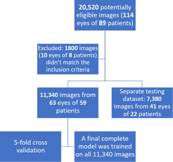
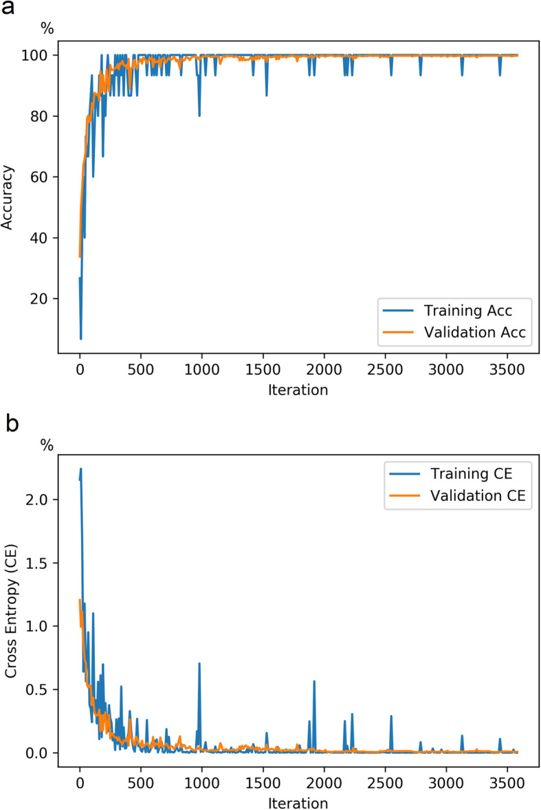
Methods: In this observational case-control study, 104 eyes (53 FECD eyes and 51 healthy controls) received HD-OCT imaging (Envisu R2210, Bioptigen, Buffalo Grove, IL, USA) using a 6 mm radial scan pattern centered on the corneal vertex. FECD was clinically categorized into early (without corneal edema) and late-stage (with corneal edema). A total of 18,720 anterior segment optical coherence tomography (AS-OCT) images (9180 healthy; 5400 early-stage FECD; 4140 late-stage FECD) of 104 eyes (81 patients) were used to develop and validate a deep learning classification network to differentiate early-stage FECD eyes from healthy eyes and those with clinical edema. Using 5-fold cross-validation on the dataset containing 11,340 OCT images (63 eyes), the network was trained with 80% of these images (3420 healthy; 3060 early-stage FECD; 2700 late-stage FECD), then tested with 20% (720 healthy; 720 early-stage FECD; 720 late-stage FECD). Thereafter, a final model was trained with the entire dataset consisting the 11,340 images and validated with a remaining 7380 images of unseen AS-OCT scans of 41 eyes (5040 healthy; 1620 early-stage FECD 720 late-stage FECD). Visualization of learned features was done, and area under curve (AUC), specificity, and sensitivity of the prediction outputs for healthy, early and late-stage FECD were computed.
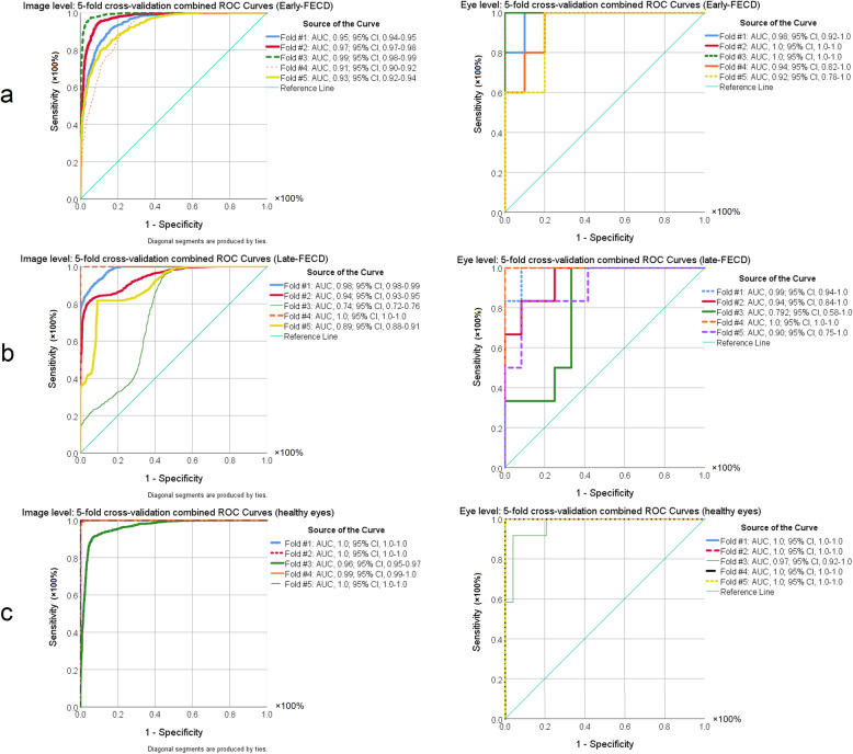
Results: The final model achieved an AUC of 0.997 ± 0.005 with 91% sensitivity and 97% specificity in detecting early-FECD; an AUC of 0.974 ± 0.005 with a specificity of 92% and a sensitivity up to 100% in detecting late-stage FECD; and an AUC of 0.998 ± 0.001 with a specificity 98% and a sensitivity of 99% in discriminating healthy corneas from all FECD.
Conclusion: Deep learning algorithm is an accurate autonomous novel diagnostic tool of FECD with very high sensitivity and specificity that can be used to grade FECD severity with high accuracy.
背景:描述一种深度学习算法在使用高清光学相干断层扫描(HD-OCT)区分无临床明显角膜水肿的早期Fuchs内皮性角膜营养不良(FECD)与健康和晚期FECD眼睛中的诊断性能。方法:在这项观察性病例对照研究中,104只眼(53只FECD眼和51只健康对照)接受了以角膜顶点为中心的6mm径向扫描模式的HD-OCT成像(Envisu R2210, Bioptigen, Buffalo Grove, IL, USA)。FECD临床分为早期(无角膜水肿)和晚期(有角膜水肿)。共18720张前段光学相干断层扫描(AS-OCT)图像(9180张健康图像;5400早期FECD;4140只晚期FECD的104只眼睛(81名患者)被用于开发和验证一个深度学习分类网络,以区分早期FECD眼睛与健康眼睛和临床水肿眼睛。对包含11,340张OCT图像(63只眼睛)的数据集使用5倍交叉验证,该网络使用80%的这些图像(3420张健康图像;3060早期FECD;2700例晚期FECD患者,然后用20%(720例健康;720早期FECD;720后期FECD)。之后,使用包含11,340张图像的整个数据集训练最终模型,并使用41只眼睛(5040只健康眼睛;1620早期FECD 720后期FECD)。对学习到的特征进行可视化,并计算健康、早期和晚期FECD预测输出的曲线下面积(AUC)、特异性和敏感性。结果:最终模型检测早期fecd的AUC为0.997±0.005,灵敏度91%,特异性97%;检测晚期FECD的AUC为0.974±0.005,特异性为92%,灵敏度高达100%;AUC为0.998±0.001,特异性为98%,灵敏度为99%。结论:深度学习算法是一种准确的FECD自主新型诊断工具,具有很高的灵敏度和特异性,可用于FECD严重程度的分级,准确率较高。

 求助内容:
求助内容: 应助结果提醒方式:


