Elodie Martin, Marie-Stéphane Aigrot, Roland Grenningloh, Bruno Stankoff, Catherine Lubetzki, Ursula Boschert, Bernard Zalc
{"title":"Bruton's Tyrosine Kinase Inhibition Promotes Myelin Repair.","authors":"Elodie Martin, Marie-Stéphane Aigrot, Roland Grenningloh, Bruno Stankoff, Catherine Lubetzki, Ursula Boschert, Bernard Zalc","doi":"10.3233/BPL-200100","DOIUrl":null,"url":null,"abstract":"<p><strong>Background: </strong>Microglia are the resident macrophages of the central nervous system (CNS). In multiple sclerosis (MS) and related experimental models, microglia have either a pro-inflammatory or a pro-regenerative/pro-remyelinating function. Inhibition of Bruton's tyrosine kinase (BTK), a member of the Tec family of kinases, has been shown to block differentiation of pro-inflammatory macrophages in response to granulocyte-macrophage colony-stimulating factor <i>in vitro</i>. However, the role of BTK in the CNS is unknown.</p><p><strong>Methods: </strong>Our aim was to investigate the effect of BTK inhibition on myelin repair in <i>ex vivo</i> and <i>in vivo</i> experimental models of demyelination and remyelination. The remyelination effect of a BTK inhibitor (BTKi; BTKi-1) was then investigated in LPC-induced demyelinated cerebellar organotypic slice cultures and metronidazole-induced demyelinated <i>Xenopus MBP-GFP-NTR</i> transgenic tadpoles.</p><p><strong>Results: </strong>Cellular detection of BTK and its activated form BTK-phospho-Y223 (p-BTK) was determined by immunohistochemistry in organotypic cerebellar slice cultures, before and after lysophosphatidylcholine (LPC)-induced demyelination. A low BTK signal detected by immunolabeling under normal conditions in cerebellar slices was in sharp contrast to an 8.5-fold increase in the number of BTK-positive cells observed in LPC-demyelinated slice cultures. Under both conditions, approximately 75% of cells expressing BTK and p-BTK were microglia and 25% were astrocytes. Compared with spontaneous recovery, treatment of demyelinated slice cultures and MTZ-demyelinated transgenic tadpoles with BTKi resulted in at least a 1.7-fold improvement of remyelination.</p><p><strong>Conclusion: </strong>Our data demonstrate that BTK inhibition is a promising therapeutic strategy for myelin repair.</p>","PeriodicalId":72451,"journal":{"name":"Brain plasticity (Amsterdam, Netherlands)","volume":"5 2","pages":"123-133"},"PeriodicalIF":0.0000,"publicationDate":"2020-10-01","publicationTypes":"Journal Article","fieldsOfStudy":null,"isOpenAccess":false,"openAccessPdf":"https://sci-hub-pdf.com/10.3233/BPL-200100","citationCount":"39","resultStr":null,"platform":"Semanticscholar","paperid":null,"PeriodicalName":"Brain plasticity (Amsterdam, Netherlands)","FirstCategoryId":"1085","ListUrlMain":"https://doi.org/10.3233/BPL-200100","RegionNum":0,"RegionCategory":null,"ArticlePicture":[],"TitleCN":null,"AbstractTextCN":null,"PMCID":null,"EPubDate":"","PubModel":"","JCR":"","JCRName":"","Score":null,"Total":0}
引用次数: 39
Abstract
Background: Microglia are the resident macrophages of the central nervous system (CNS). In multiple sclerosis (MS) and related experimental models, microglia have either a pro-inflammatory or a pro-regenerative/pro-remyelinating function. Inhibition of Bruton's tyrosine kinase (BTK), a member of the Tec family of kinases, has been shown to block differentiation of pro-inflammatory macrophages in response to granulocyte-macrophage colony-stimulating factor in vitro. However, the role of BTK in the CNS is unknown.
Methods: Our aim was to investigate the effect of BTK inhibition on myelin repair in ex vivo and in vivo experimental models of demyelination and remyelination. The remyelination effect of a BTK inhibitor (BTKi; BTKi-1) was then investigated in LPC-induced demyelinated cerebellar organotypic slice cultures and metronidazole-induced demyelinated Xenopus MBP-GFP-NTR transgenic tadpoles.
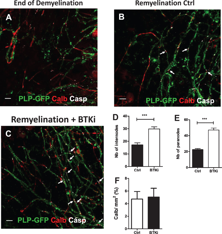
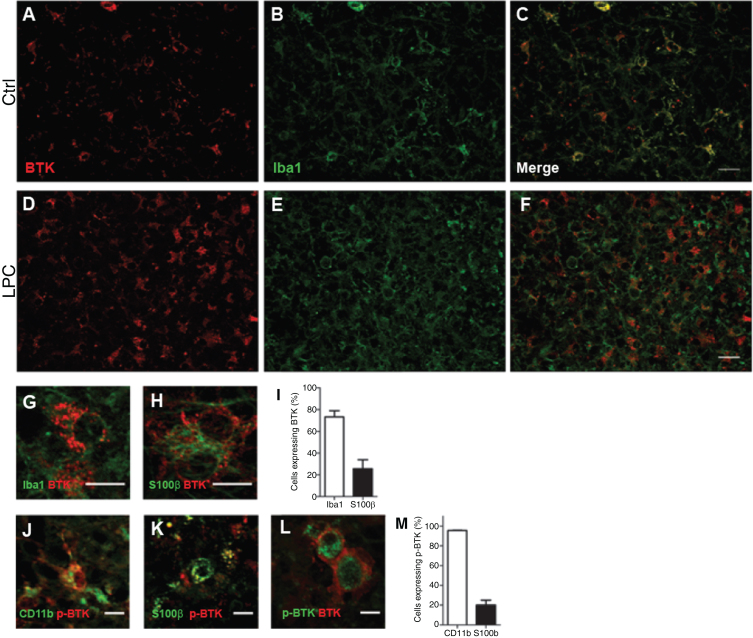
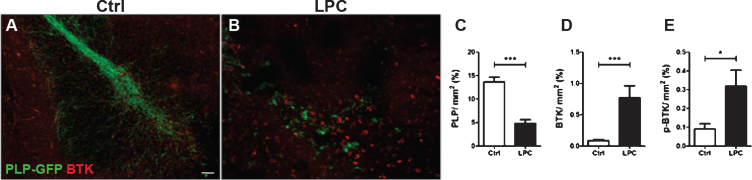
Results: Cellular detection of BTK and its activated form BTK-phospho-Y223 (p-BTK) was determined by immunohistochemistry in organotypic cerebellar slice cultures, before and after lysophosphatidylcholine (LPC)-induced demyelination. A low BTK signal detected by immunolabeling under normal conditions in cerebellar slices was in sharp contrast to an 8.5-fold increase in the number of BTK-positive cells observed in LPC-demyelinated slice cultures. Under both conditions, approximately 75% of cells expressing BTK and p-BTK were microglia and 25% were astrocytes. Compared with spontaneous recovery, treatment of demyelinated slice cultures and MTZ-demyelinated transgenic tadpoles with BTKi resulted in at least a 1.7-fold improvement of remyelination.
Conclusion: Our data demonstrate that BTK inhibition is a promising therapeutic strategy for myelin repair.

 求助内容:
求助内容: 应助结果提醒方式:


