{"title":"Evaluation of plantar fascia using high-resolution ultrasonography in clinically diagnosed cases of plantar fasciitis.","authors":"Purnima Aggarwal, Vivek Jirankali, Sudhir K Garg","doi":"10.5114/pjr.2020.97955","DOIUrl":null,"url":null,"abstract":"<p><strong>Purpose: </strong>The aim of this study was to assess the efficacy of high-resolution ultrasonography in the assessment of plantar fascia in individuals with heel pain, before and after treatment.</p><p><strong>Material and methods: </strong>This study was conducted from 2016 to 2019, during which time 44 clinically diagnosed patients of plantar fasciitis were compared to 50 normal volunteers. There were 25 males and 25 females in the control group and 42 females and two males in the study group. Thirty-eight patients had unilateral disease, and six patients had bilateral disease. The thickness of the plantar fascia was measured just anterior to its calcaneal attachment using ultrasonography. Body mass index (BMI) was also calculated in both groups.</p><p><strong>Results: </strong>The plantar fascia was 2-4 mm thick in the control group whereas it was > 4 mm thick in 48 heels in the study group. With cut-off of > 4 mm as diagnostic of plantar fasciitis, this study had a sensitivity of 96%, specificity of 100%, and accuracy of 98%. BMI was increased in 60% of female patients. All patients were treated with local infiltration of corticosteroid. In 37/42 patients (43 heels) who had improved clinically, the thickness of plantar fascia was reduced to < 4 mm when assessed after six weeks of corticosteroid injection.</p><p><strong>Conclusions: </strong>Diagnosis of plantar fasciitis can be easily verified by ultrasonography with plantar fascia thickness > 4 mm being suggestive of plantar fasciitis. Ultrasound can also be used to evaluate treatment response. Ultrasono-graphy helps the clinician in confirming the diagnosis of plantar fasciitis and also in assessing the response to treatment.</p>","PeriodicalId":47128,"journal":{"name":"Polish Journal of Radiology","volume":"85 ","pages":"e375-e380"},"PeriodicalIF":0.9000,"publicationDate":"2020-07-24","publicationTypes":"Journal Article","fieldsOfStudy":null,"isOpenAccess":false,"openAccessPdf":"https://ftp.ncbi.nlm.nih.gov/pub/pmc/oa_pdf/5e/9e/PJR-85-41528.PMC7425221.pdf","citationCount":"13","resultStr":null,"platform":"Semanticscholar","paperid":null,"PeriodicalName":"Polish Journal of Radiology","FirstCategoryId":"1085","ListUrlMain":"https://doi.org/10.5114/pjr.2020.97955","RegionNum":0,"RegionCategory":null,"ArticlePicture":[],"TitleCN":null,"AbstractTextCN":null,"PMCID":null,"EPubDate":"2020/1/1 0:00:00","PubModel":"eCollection","JCR":"Q4","JCRName":"RADIOLOGY, NUCLEAR MEDICINE & MEDICAL IMAGING","Score":null,"Total":0}
引用次数: 13
Abstract
Purpose: The aim of this study was to assess the efficacy of high-resolution ultrasonography in the assessment of plantar fascia in individuals with heel pain, before and after treatment.
Material and methods: This study was conducted from 2016 to 2019, during which time 44 clinically diagnosed patients of plantar fasciitis were compared to 50 normal volunteers. There were 25 males and 25 females in the control group and 42 females and two males in the study group. Thirty-eight patients had unilateral disease, and six patients had bilateral disease. The thickness of the plantar fascia was measured just anterior to its calcaneal attachment using ultrasonography. Body mass index (BMI) was also calculated in both groups.
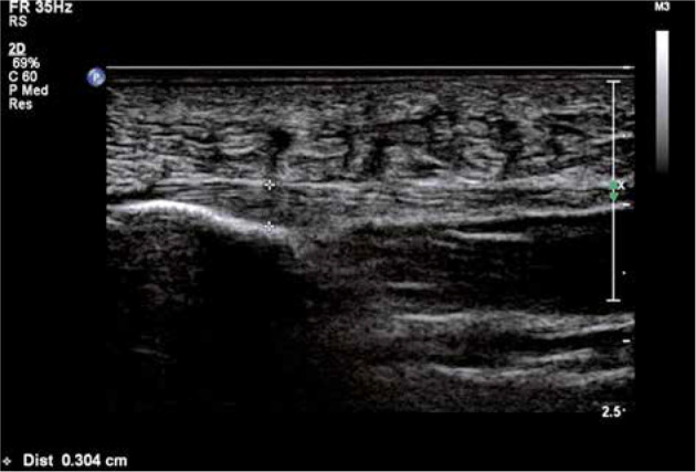
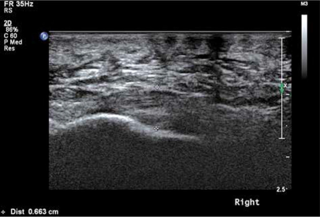
Results: The plantar fascia was 2-4 mm thick in the control group whereas it was > 4 mm thick in 48 heels in the study group. With cut-off of > 4 mm as diagnostic of plantar fasciitis, this study had a sensitivity of 96%, specificity of 100%, and accuracy of 98%. BMI was increased in 60% of female patients. All patients were treated with local infiltration of corticosteroid. In 37/42 patients (43 heels) who had improved clinically, the thickness of plantar fascia was reduced to < 4 mm when assessed after six weeks of corticosteroid injection.
Conclusions: Diagnosis of plantar fasciitis can be easily verified by ultrasonography with plantar fascia thickness > 4 mm being suggestive of plantar fasciitis. Ultrasound can also be used to evaluate treatment response. Ultrasono-graphy helps the clinician in confirming the diagnosis of plantar fasciitis and also in assessing the response to treatment.


 求助内容:
求助内容: 应助结果提醒方式:


