{"title":"Cone beam computed tomography - extending its arm to explore new possibilities.","authors":"Prajakta P Chaudhari, Chetan Bhadage, Ajay Bhoosreddy, Pragati Bramhe, Prutha Rathod, Shreya Dange","doi":"10.5114/pjr.2020.97656","DOIUrl":null,"url":null,"abstract":"<p><strong>Purpose: </strong>To determine whether the greyscale value depicted in cone beam computed tomography (CBCT) differentiates different benign osseous lesions of jaws and to measure the greyscale value of various osseous lesions of jaws and to find the correlation (if any) of these greyscale values to that of histopathological diagnosis.</p><p><strong>Material and methods: </strong>This study was conducted in the Department of Oral Medicine and Radiology of the Dental Institute after obtaining approval from the Ethical Committee. CBCT scans of osseous lesions of jaws confirmed with histopathological reports depicting cystic or tumour-like lesions were included in the study. Greyscale values depicted in CBCT scans of osseous lesions were measured. The greyscale values were grouped as per the histopathological diagnosis, and these ranges were then tabulated and statistically evaluated.</p><p><strong>Results: </strong>The mean value with standard deviation of greyscale values for cystic lesions was 1208.375 ± 93 and for that of the tumour group was 1603 ± 425.5.</p><p><strong>Conclusions: </strong>The greyscale value is a useful tool in differentiating between different groups of osseous lesions of jaws.</p>","PeriodicalId":47128,"journal":{"name":"Polish Journal of Radiology","volume":"85 ","pages":"e348-e352"},"PeriodicalIF":0.9000,"publicationDate":"2020-07-10","publicationTypes":"Journal Article","fieldsOfStudy":null,"isOpenAccess":false,"openAccessPdf":"https://ftp.ncbi.nlm.nih.gov/pub/pmc/oa_pdf/46/91/PJR-85-41424.PMC7425220.pdf","citationCount":"0","resultStr":null,"platform":"Semanticscholar","paperid":null,"PeriodicalName":"Polish Journal of Radiology","FirstCategoryId":"1085","ListUrlMain":"https://doi.org/10.5114/pjr.2020.97656","RegionNum":0,"RegionCategory":null,"ArticlePicture":[],"TitleCN":null,"AbstractTextCN":null,"PMCID":null,"EPubDate":"2020/1/1 0:00:00","PubModel":"eCollection","JCR":"Q4","JCRName":"RADIOLOGY, NUCLEAR MEDICINE & MEDICAL IMAGING","Score":null,"Total":0}
引用次数: 0
Abstract
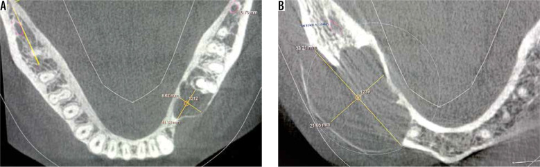
Purpose: To determine whether the greyscale value depicted in cone beam computed tomography (CBCT) differentiates different benign osseous lesions of jaws and to measure the greyscale value of various osseous lesions of jaws and to find the correlation (if any) of these greyscale values to that of histopathological diagnosis.
Material and methods: This study was conducted in the Department of Oral Medicine and Radiology of the Dental Institute after obtaining approval from the Ethical Committee. CBCT scans of osseous lesions of jaws confirmed with histopathological reports depicting cystic or tumour-like lesions were included in the study. Greyscale values depicted in CBCT scans of osseous lesions were measured. The greyscale values were grouped as per the histopathological diagnosis, and these ranges were then tabulated and statistically evaluated.
Results: The mean value with standard deviation of greyscale values for cystic lesions was 1208.375 ± 93 and for that of the tumour group was 1603 ± 425.5.
Conclusions: The greyscale value is a useful tool in differentiating between different groups of osseous lesions of jaws.

 求助内容:
求助内容: 应助结果提醒方式:


