{"title":"Calcifying Fibrous Tumor of the Mesentery: A Case Report and a Review of the Literature.","authors":"Derqaoui Sabrine, Elouazzani Hafsa, Ratbi Amine, Bernoussi Zakia, Zouaidia Fouad","doi":"10.1177/2632010X20930689","DOIUrl":null,"url":null,"abstract":"<p><strong>Background: </strong>Calcifying fibrous tumor (CFT) is a rare entity, with a distinctive histological presentation, initially reported as childhood fibrous tumor with psammoma bodies. It is a benign hypocellular fibrous neoplasm calcifications and lymphoplasmacytic infiltrate. The CFTs may involve many sites, including gastrointestinal tract, pleura, abdominal cavity, and neck. The diagnosis might be challenging due to histological overlaps with other mesenchymal tumors. The prognosis is good. We describe herein the case of a 53-year-old woman with an incidentally diagnosed CFT of the mesentery.</p><p><strong>Case presentation: </strong>A 53-year-old woman presented to the surgery department with a 2-year history of an anterior abdominal hernia. A computed tomographic scan of the abdomen failed to demonstrate any evidence of a mesenteric nodule. The patient underwent surgical treatment. Careful exploration during the excision of herniated sac revealed a solitary nodule of the mesentery. Local excision was performed. On gross, it was a well-demarcated nodule. Microscopically, the tumor consisted of an abundant paucicellular hyalinized collagen with calcifications; associated to a sparse mononuclear inflammatory infiltrate.</p><p><strong>Conclusions: </strong>Calcifying fibrous tumor is a benign lesion. The diagnosis is based on histology, because clinical and radiological features are nonspecific. Awareness of this entity is crucial to distinguish it from other mesenchymal tumors especially in the gastrointestinal tract.</p>","PeriodicalId":53204,"journal":{"name":"Clinical Pathology","volume":"13 ","pages":"2632010X20930689"},"PeriodicalIF":1.9000,"publicationDate":"2020-06-23","publicationTypes":"Journal Article","fieldsOfStudy":null,"isOpenAccess":false,"openAccessPdf":"https://sci-hub-pdf.com/10.1177/2632010X20930689","citationCount":"0","resultStr":null,"platform":"Semanticscholar","paperid":null,"PeriodicalName":"Clinical Pathology","FirstCategoryId":"1085","ListUrlMain":"https://doi.org/10.1177/2632010X20930689","RegionNum":0,"RegionCategory":null,"ArticlePicture":[],"TitleCN":null,"AbstractTextCN":null,"PMCID":null,"EPubDate":"2020/1/1 0:00:00","PubModel":"eCollection","JCR":"Q3","JCRName":"PATHOLOGY","Score":null,"Total":0}
引用次数: 0
Abstract
Background: Calcifying fibrous tumor (CFT) is a rare entity, with a distinctive histological presentation, initially reported as childhood fibrous tumor with psammoma bodies. It is a benign hypocellular fibrous neoplasm calcifications and lymphoplasmacytic infiltrate. The CFTs may involve many sites, including gastrointestinal tract, pleura, abdominal cavity, and neck. The diagnosis might be challenging due to histological overlaps with other mesenchymal tumors. The prognosis is good. We describe herein the case of a 53-year-old woman with an incidentally diagnosed CFT of the mesentery.
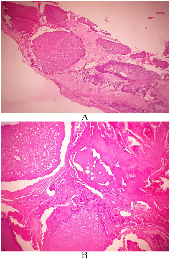
Case presentation: A 53-year-old woman presented to the surgery department with a 2-year history of an anterior abdominal hernia. A computed tomographic scan of the abdomen failed to demonstrate any evidence of a mesenteric nodule. The patient underwent surgical treatment. Careful exploration during the excision of herniated sac revealed a solitary nodule of the mesentery. Local excision was performed. On gross, it was a well-demarcated nodule. Microscopically, the tumor consisted of an abundant paucicellular hyalinized collagen with calcifications; associated to a sparse mononuclear inflammatory infiltrate.
Conclusions: Calcifying fibrous tumor is a benign lesion. The diagnosis is based on histology, because clinical and radiological features are nonspecific. Awareness of this entity is crucial to distinguish it from other mesenchymal tumors especially in the gastrointestinal tract.


 求助内容:
求助内容: 应助结果提醒方式:


