Matthew B Greenberg, Nandini Venkateswaran, Ann Q Tran, Ranya G Habash, Wendy W Lee
{"title":"Vision Loss Secondary to Meningioma in the Pregnant Patient.","authors":"Matthew B Greenberg, Nandini Venkateswaran, Ann Q Tran, Ranya G Habash, Wendy W Lee","doi":"","DOIUrl":null,"url":null,"abstract":"<p><p>A 31-year-old primagravid female at 27 weeks gestation presented to the emergency room with three weeks of progressive blurring of vision associated with intermittent headaches. Ocular examination revealed diminished visual acuity, decreased color discrimination, and constricted confrontation visual fields; optic nerve appearance was however normal. Magnetic resonance imaging of the brain and orbits revealed a large tuberculum meningioma compressing the optic chiasm and prechiasmatic optic nerves, as well as a small sphenoid wing meningioma. Given the risk of permanent vision loss, the patient underwent emergent tumor resection. Near total resection of the masses was achieved and the patient had complete resolution of her vision post-operatively. She gave birth via Caesarean section at 39 weeks. This case report describes the clinical presentations of intracranial meningiomas and discusses the challenges this condition poses in management during pregnancy.</p>","PeriodicalId":93021,"journal":{"name":"Journal of clinical ophthalmology & eye disorders","volume":"1 1","pages":""},"PeriodicalIF":0.0000,"publicationDate":"2017-01-01","publicationTypes":"Journal Article","fieldsOfStudy":null,"isOpenAccess":false,"openAccessPdf":"https://www.ncbi.nlm.nih.gov/pmc/articles/PMC7241594/pdf/","citationCount":"0","resultStr":null,"platform":"Semanticscholar","paperid":null,"PeriodicalName":"Journal of clinical ophthalmology & eye disorders","FirstCategoryId":"1085","ListUrlMain":"","RegionNum":0,"RegionCategory":null,"ArticlePicture":[],"TitleCN":null,"AbstractTextCN":null,"PMCID":null,"EPubDate":"2017/9/11 0:00:00","PubModel":"Epub","JCR":"","JCRName":"","Score":null,"Total":0}
引用次数: 0
Abstract
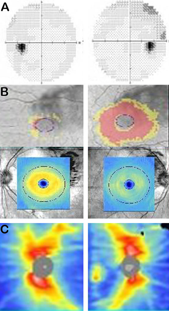
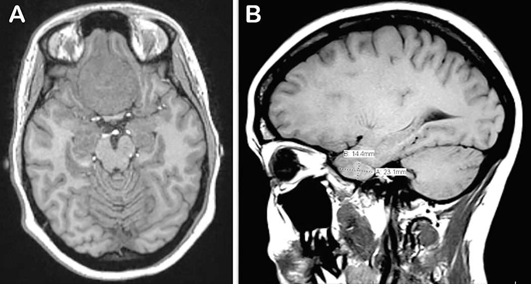
A 31-year-old primagravid female at 27 weeks gestation presented to the emergency room with three weeks of progressive blurring of vision associated with intermittent headaches. Ocular examination revealed diminished visual acuity, decreased color discrimination, and constricted confrontation visual fields; optic nerve appearance was however normal. Magnetic resonance imaging of the brain and orbits revealed a large tuberculum meningioma compressing the optic chiasm and prechiasmatic optic nerves, as well as a small sphenoid wing meningioma. Given the risk of permanent vision loss, the patient underwent emergent tumor resection. Near total resection of the masses was achieved and the patient had complete resolution of her vision post-operatively. She gave birth via Caesarean section at 39 weeks. This case report describes the clinical presentations of intracranial meningiomas and discusses the challenges this condition poses in management during pregnancy.

 求助内容:
求助内容: 应助结果提醒方式:


