{"title":"Detection of LC3-Associated Phagocytosis (LAP)","authors":"Jennifer Martinez","doi":"10.1002/cpcb.104","DOIUrl":null,"url":null,"abstract":"<p>Phagocytes, notably macrophages, are critical sentinels of their environment, patrolling for and eradicating unwanted components. The ability of cells to process extracellular cargo in an appropriate manner is important for both clearance of the cargo and eventual return to homeostasis. Although the evolutionarily conserved pathway of autophagy involves the degradation and recycling of unnecessary or dysfunctional cellular components during starvation, we now appreciate that the reach of autophagy extends beyond nutrient deprivation, notably including cellular quality control (e.g., mitophagy) and host defense against internalized pathogens (i.e., xenophagy). Despite being seemingly disparate, autophagic functions are unified as conserved mechanisms for containment and immunosuppression, suggesting an original immune function for autophagy. A recently described pathway called LC3-associated phagocytosis (LAP) marries the ancient concepts of phagocytosis and autophagy, revealing new ways in which the autophagy machinery, in a molecularly distinct pathway, contributes to the inflammatory response. In this article, protocols to detect LAP by electron microscopy, immunofluorescence, flow cytometry, and phagosome purification are described, allowing the user to detect multiple characteristics of LAP in both qualitative and quantitative manners. Published 2020. U.S. Government.</p><p><b>Basic Protocol 1</b>: Detection of LAP by electron microscopy</p><p><b>Basic Protocol 2</b>: Detection of LAP by confocal microscopy of LC3-GFP-expressing cells</p><p><b>Alternate Protocol 1</b>: Detection of LAP by confocal microscopy using immunofluorescence</p><p><b>Basic Protocol 3</b>: Detection of LAP using flow cytometry of LC3-GFP-expressing cells</p><p><b>Alternate Protocol 2</b>: Detection of LAP using antibody staining and flow cytometry</p><p><b>Basic Protocol 4</b>: Detection of LAP by western blot of purified LAPosomes</p>","PeriodicalId":40051,"journal":{"name":"Current Protocols in Cell Biology","volume":"87 1","pages":""},"PeriodicalIF":0.0000,"publicationDate":"2020-05-21","publicationTypes":"Journal Article","fieldsOfStudy":null,"isOpenAccess":false,"openAccessPdf":"https://sci-hub-pdf.com/10.1002/cpcb.104","citationCount":"11","resultStr":null,"platform":"Semanticscholar","paperid":null,"PeriodicalName":"Current Protocols in Cell Biology","FirstCategoryId":"1085","ListUrlMain":"https://onlinelibrary.wiley.com/doi/10.1002/cpcb.104","RegionNum":0,"RegionCategory":null,"ArticlePicture":[],"TitleCN":null,"AbstractTextCN":null,"PMCID":null,"EPubDate":"","PubModel":"","JCR":"Q3","JCRName":"Biochemistry, Genetics and Molecular Biology","Score":null,"Total":0}
引用次数: 11
Abstract
Phagocytes, notably macrophages, are critical sentinels of their environment, patrolling for and eradicating unwanted components. The ability of cells to process extracellular cargo in an appropriate manner is important for both clearance of the cargo and eventual return to homeostasis. Although the evolutionarily conserved pathway of autophagy involves the degradation and recycling of unnecessary or dysfunctional cellular components during starvation, we now appreciate that the reach of autophagy extends beyond nutrient deprivation, notably including cellular quality control (e.g., mitophagy) and host defense against internalized pathogens (i.e., xenophagy). Despite being seemingly disparate, autophagic functions are unified as conserved mechanisms for containment and immunosuppression, suggesting an original immune function for autophagy. A recently described pathway called LC3-associated phagocytosis (LAP) marries the ancient concepts of phagocytosis and autophagy, revealing new ways in which the autophagy machinery, in a molecularly distinct pathway, contributes to the inflammatory response. In this article, protocols to detect LAP by electron microscopy, immunofluorescence, flow cytometry, and phagosome purification are described, allowing the user to detect multiple characteristics of LAP in both qualitative and quantitative manners. Published 2020. U.S. Government.
Basic Protocol 1: Detection of LAP by electron microscopy
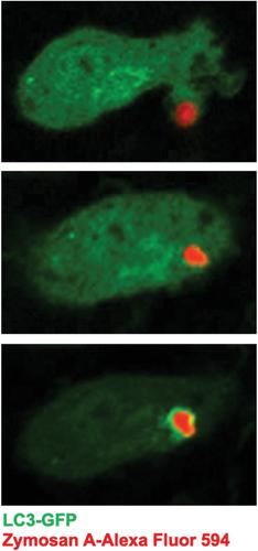
Basic Protocol 2: Detection of LAP by confocal microscopy of LC3-GFP-expressing cells
Alternate Protocol 1: Detection of LAP by confocal microscopy using immunofluorescence
Basic Protocol 3: Detection of LAP using flow cytometry of LC3-GFP-expressing cells
Alternate Protocol 2: Detection of LAP using antibody staining and flow cytometry
Basic Protocol 4: Detection of LAP by western blot of purified LAPosomes
期刊介绍:
Developed by leading scientists in the field, Current Protocols in Cell Biology is an essential reference for researchers who study the relationship between specific molecules and genes and their location, function and structure at the cellular level. Updated every three months in all formats, CPCB is constantly evolving to keep pace with the very latest discoveries and developments.

 求助内容:
求助内容: 应助结果提醒方式:


