Carly A Clark, Cameron P Worden, Brian D Thorp, Charles S Ebert, Adam M Zanation, Brent A Senior, Steven M Johnson, Wade G McClain, Adam J Kimple
{"title":"Extramedullary Hematopoiesis in the Sinonasal Cavity: A Case Report and Review of the Literature.","authors":"Carly A Clark, Cameron P Worden, Brian D Thorp, Charles S Ebert, Adam M Zanation, Brent A Senior, Steven M Johnson, Wade G McClain, Adam J Kimple","doi":"10.1177/2152656720918874","DOIUrl":null,"url":null,"abstract":"<p><strong>Background: </strong>Extramedullary hematopoiesis (EMH) occurs in patients with hematologic disorders, but rarely within the paranasal sinuses. We report a case of EMH in a 17-year-old male with sickle cell disease (SCD) who presented with occipital pain and sinusitis. A computed tomography (CT) scan demonstrated heterogeneous opacification of the right maxillary sinus concerning for allergic fungal sinusitis or a fungal ball with bony erosion. He was taken to the operating room for endoscopic biopsy and a limited endoscopic sinus surgery. Grossly, his maxillary sinus was filled with spiculated osseous tissue. Final pathology demonstrated active hematopoietic bone marrow filling the sinus.</p><p><strong>Methods: </strong>We present a case report and literature review of sinonasal EMH.</p><p><strong>Results: </strong>We identified 14 articles with 15 patients. EMH was typically associated with SCD or beta thalassemia. The average age of presentation was 30. There was a male sex predilection with a ratio of 11:15. The most common presenting symptom was a headache and nasal obstruction (33% for both). The most common finding on CT was a soft tissue expansile mass (73%). The most commonly affected location was the maxillary sinus (60%).</p><p><strong>Conclusions: </strong>This case report serves as a reminder to consider EMH as an uncommon cause of sinus opacification, particularly in patients with SCD or beta thalassemia. The expansion of hematopoietic tissue may be identified as a sinus mass on CT. By recognizing the potential manifestations of chronic anemia, an accurate and timely diagnosis can be made.</p>","PeriodicalId":45192,"journal":{"name":"Allergy & Rhinology","volume":" ","pages":"2152656720918874"},"PeriodicalIF":1.2000,"publicationDate":"2020-04-21","publicationTypes":"Journal Article","fieldsOfStudy":null,"isOpenAccess":false,"openAccessPdf":"https://sci-hub-pdf.com/10.1177/2152656720918874","citationCount":"2","resultStr":null,"platform":"Semanticscholar","paperid":null,"PeriodicalName":"Allergy & Rhinology","FirstCategoryId":"1085","ListUrlMain":"https://doi.org/10.1177/2152656720918874","RegionNum":0,"RegionCategory":null,"ArticlePicture":[],"TitleCN":null,"AbstractTextCN":null,"PMCID":null,"EPubDate":"2020/1/1 0:00:00","PubModel":"eCollection","JCR":"Q1","JCRName":"OTORHINOLARYNGOLOGY","Score":null,"Total":0}
引用次数: 2
Abstract
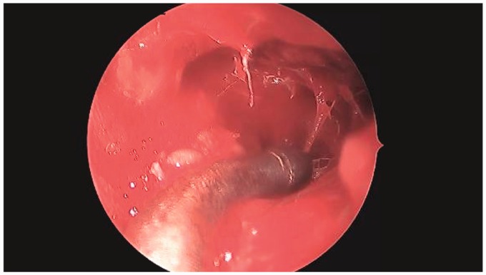
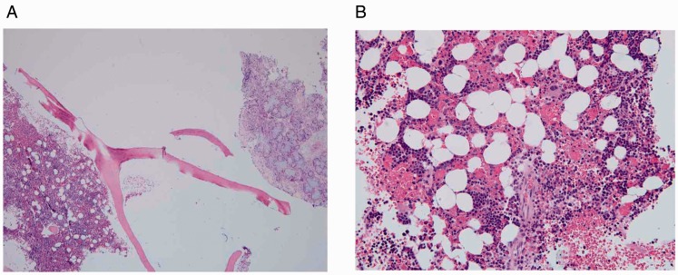
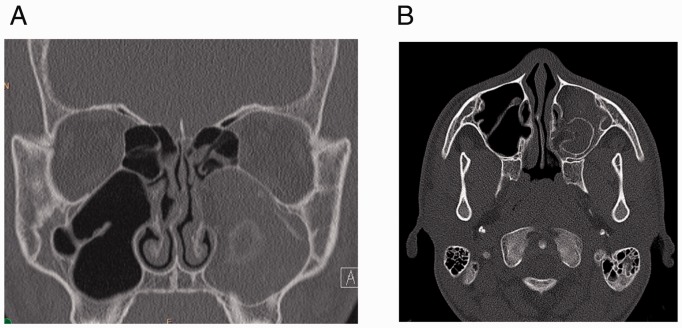
Background: Extramedullary hematopoiesis (EMH) occurs in patients with hematologic disorders, but rarely within the paranasal sinuses. We report a case of EMH in a 17-year-old male with sickle cell disease (SCD) who presented with occipital pain and sinusitis. A computed tomography (CT) scan demonstrated heterogeneous opacification of the right maxillary sinus concerning for allergic fungal sinusitis or a fungal ball with bony erosion. He was taken to the operating room for endoscopic biopsy and a limited endoscopic sinus surgery. Grossly, his maxillary sinus was filled with spiculated osseous tissue. Final pathology demonstrated active hematopoietic bone marrow filling the sinus.
Methods: We present a case report and literature review of sinonasal EMH.
Results: We identified 14 articles with 15 patients. EMH was typically associated with SCD or beta thalassemia. The average age of presentation was 30. There was a male sex predilection with a ratio of 11:15. The most common presenting symptom was a headache and nasal obstruction (33% for both). The most common finding on CT was a soft tissue expansile mass (73%). The most commonly affected location was the maxillary sinus (60%).
Conclusions: This case report serves as a reminder to consider EMH as an uncommon cause of sinus opacification, particularly in patients with SCD or beta thalassemia. The expansion of hematopoietic tissue may be identified as a sinus mass on CT. By recognizing the potential manifestations of chronic anemia, an accurate and timely diagnosis can be made.

 求助内容:
求助内容: 应助结果提醒方式:


