{"title":"Reduced macular inner retinal thickness and microvascular density in the early stage of patients with dysthyroid optic neuropathy.","authors":"Yufei Wu, Yunhai Tu, Chaoming Wu, Lulu Bao, Jianhua Wang, Fan Lu, Meixiao Shen, Qi Chen","doi":"10.1186/s40662-020-00180-9","DOIUrl":null,"url":null,"abstract":"<p><strong>Background: </strong>The goal was to investigate changes of the inner intra-retinal layer thicknesses and retinal capillary density (RCD) around the macula in thyroid-associated ophthalmopathy (TAO) patients with or without dysthyroid optic neuropathy (DON).</p><p><strong>Methods: </strong>Forty-four TAO patients including 23 non-DON and 21 DON patients, and 38 healthy participants were enrolled. Spectral domain optical coherence tomography equipped with Angiovue was used to obtain three-dimensional retinal thickness maps and microvascular images of the superficial and deep retinal capillary layers (SRCL and DRCL, respectively) around the macula. Quantitative analyses were performed using a custom automated algorithm.</p><p><strong>Results: </strong>The thicknesses of the nerve fiber layer, ganglion cell layer + inner plexiform layer, and ganglion cell complex (GCC) as well as the RCDs in the SRCL and DRCL in both TAO groups were significantly decreased compared to the controls. In addition, the RCDs in DRCL of the DON group were further decreased compared to the non-DON group. GCC thickness in both TAO groups was positively correlated with the RCDs of the SRCL in the total annular zone and in the temporal, inferior, and nasal sectors. The areas under the receiver operating characteristic curves for the GCC thickness combined with the RCD were generally larger than those of each single indicator.</p><p><strong>Conclusions: </strong>Thinned inner intra-retinal layers and decreased RCDs in the TAO patients without DON revealed that morphological changes might precede visual dysfunction. The composite index of the retinal structure and the microvascular density might be valuable in the diagnosing, monitoring, and intervention for early DON.</p>","PeriodicalId":520624,"journal":{"name":"Eye and vision (London, England)","volume":" ","pages":"16"},"PeriodicalIF":4.0000,"publicationDate":"2020-03-10","publicationTypes":"Journal Article","fieldsOfStudy":null,"isOpenAccess":false,"openAccessPdf":"https://sci-hub-pdf.com/10.1186/s40662-020-00180-9","citationCount":"24","resultStr":null,"platform":"Semanticscholar","paperid":null,"PeriodicalName":"Eye and vision (London, England)","FirstCategoryId":"3","ListUrlMain":"https://doi.org/10.1186/s40662-020-00180-9","RegionNum":0,"RegionCategory":null,"ArticlePicture":[],"TitleCN":null,"AbstractTextCN":null,"PMCID":null,"EPubDate":"2020/1/1 0:00:00","PubModel":"eCollection","JCR":"","JCRName":"","Score":null,"Total":0}
引用次数: 24
Abstract
Background: The goal was to investigate changes of the inner intra-retinal layer thicknesses and retinal capillary density (RCD) around the macula in thyroid-associated ophthalmopathy (TAO) patients with or without dysthyroid optic neuropathy (DON).
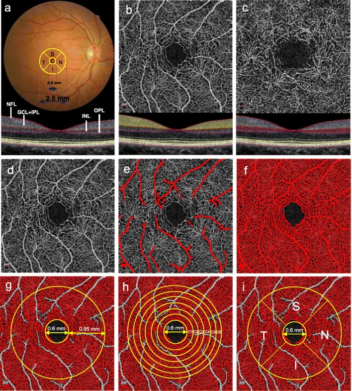
Methods: Forty-four TAO patients including 23 non-DON and 21 DON patients, and 38 healthy participants were enrolled. Spectral domain optical coherence tomography equipped with Angiovue was used to obtain three-dimensional retinal thickness maps and microvascular images of the superficial and deep retinal capillary layers (SRCL and DRCL, respectively) around the macula. Quantitative analyses were performed using a custom automated algorithm.
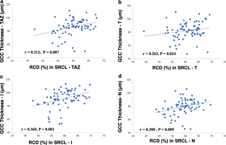
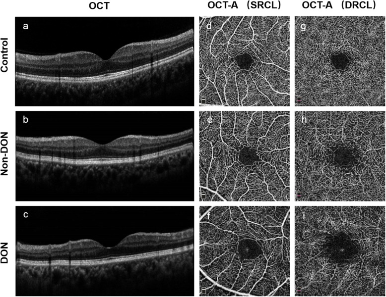
Results: The thicknesses of the nerve fiber layer, ganglion cell layer + inner plexiform layer, and ganglion cell complex (GCC) as well as the RCDs in the SRCL and DRCL in both TAO groups were significantly decreased compared to the controls. In addition, the RCDs in DRCL of the DON group were further decreased compared to the non-DON group. GCC thickness in both TAO groups was positively correlated with the RCDs of the SRCL in the total annular zone and in the temporal, inferior, and nasal sectors. The areas under the receiver operating characteristic curves for the GCC thickness combined with the RCD were generally larger than those of each single indicator.
Conclusions: Thinned inner intra-retinal layers and decreased RCDs in the TAO patients without DON revealed that morphological changes might precede visual dysfunction. The composite index of the retinal structure and the microvascular density might be valuable in the diagnosing, monitoring, and intervention for early DON.

 求助内容:
求助内容: 应助结果提醒方式:


