Mature Ovarian Teratoma: Atypical Imaging.
Case Reports in Radiology
Pub Date : 2020-02-18
eCollection Date: 2020-01-01
DOI:10.1155/2020/1352961
引用次数: 0
Abstract
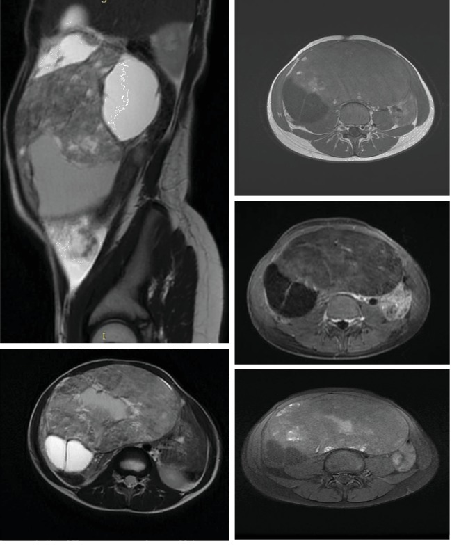
The incidence of a mature ovarian teratoma ranged from 20% to 30% of pediatric ovarian tumors (Sabaa et al., 2009), which is composed of well-differentiated tissues that derive from all three germ cell layers (ectoderm, mesoderm, and endoderm); it is one of the most common benign ovarian neoplasms. In this case report, we discuss a 9-year-old female patient who presented with abdominal pain and distended abdomen, for which she had an abdominal ultrasound and magnetic resonance imaging. The histopathological exam, after a laparotomy, showed a mature ovarian teratoma.


成熟卵巢畸胎瘤:不典型影像。
成熟卵巢畸胎瘤的发生率在儿童卵巢肿瘤的20%至30%之间(Sabaa等,2009),它由来自所有三个生殖细胞层(外胚层、中胚层和内胚层)的分化良好的组织组成;它是最常见的良性卵巢肿瘤之一。在这个病例报告中,我们讨论了一个9岁的女性患者,她表现为腹痛和腹部膨胀,为此她做了腹部超声和磁共振成像。剖腹手术后的组织病理学检查显示为成熟卵巢畸胎瘤。
本文章由计算机程序翻译,如有差异,请以英文原文为准。
求助全文
约1分钟内获得全文
求助全文

 求助内容:
求助内容: 应助结果提醒方式:


