Yeli Pi, Shilpa Radhakrishnan, Yaser Alrajhi, Ravi Bhargava
{"title":"Magnetic Apposition across the Epiglottis: Radiographic and Operative Correlation of a Rare Hypopharyngeal Foreign Body.","authors":"Yeli Pi, Shilpa Radhakrishnan, Yaser Alrajhi, Ravi Bhargava","doi":"10.1155/2020/3245634","DOIUrl":null,"url":null,"abstract":"<p><p><i>Background and Aim</i>. Rare-earth magnet ingestions are a subset of foreign body ingestions and can result in significant morbidity secondary to pressure necrosis. These magnets are best visualized radiographically, typically located in the gastrointestinal tract. However, unusual locations of magnetic adherence may include the hypopharynx along the epiglottis, where only 2 previous cases have been reported. Clinicians should be aware of the potential dangers of rare-earth magnet ingestion and consider atypical locations of attachment in the appropriate clinical setting. <i>Case Presentation</i>. We present an interesting case of a fourteen-year-old female patient who presents with witnessed ingestion of multiple rare-earth magnets. Soft-tissue neck radiographs demonstrate two adjacent rounded radiopaque densities in the hypopharynx. Intraoperative images confirmed the radiographic findings and identified two magnetic balls stuck along the dorsal and ventral aspect of the epiglottis without evidence of pressure necrosis.</p><p><strong>Conclusion: </strong>This is the first published case of magnetic foreign body adherence to the epiglottis in the Radiology literature. Awareness and recognition of the unique radiographic findings of this rare entity can help clinicians streamline timely management.</p>","PeriodicalId":30326,"journal":{"name":"Case Reports in Radiology","volume":"2020 ","pages":"3245634"},"PeriodicalIF":0.0000,"publicationDate":"2020-02-03","publicationTypes":"Journal Article","fieldsOfStudy":null,"isOpenAccess":false,"openAccessPdf":"https://sci-hub-pdf.com/10.1155/2020/3245634","citationCount":"1","resultStr":null,"platform":"Semanticscholar","paperid":null,"PeriodicalName":"Case Reports in Radiology","FirstCategoryId":"1085","ListUrlMain":"https://doi.org/10.1155/2020/3245634","RegionNum":0,"RegionCategory":null,"ArticlePicture":[],"TitleCN":null,"AbstractTextCN":null,"PMCID":null,"EPubDate":"2020/1/1 0:00:00","PubModel":"eCollection","JCR":"","JCRName":"","Score":null,"Total":0}
引用次数: 1
Abstract
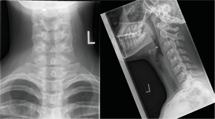
Background and Aim. Rare-earth magnet ingestions are a subset of foreign body ingestions and can result in significant morbidity secondary to pressure necrosis. These magnets are best visualized radiographically, typically located in the gastrointestinal tract. However, unusual locations of magnetic adherence may include the hypopharynx along the epiglottis, where only 2 previous cases have been reported. Clinicians should be aware of the potential dangers of rare-earth magnet ingestion and consider atypical locations of attachment in the appropriate clinical setting. Case Presentation. We present an interesting case of a fourteen-year-old female patient who presents with witnessed ingestion of multiple rare-earth magnets. Soft-tissue neck radiographs demonstrate two adjacent rounded radiopaque densities in the hypopharynx. Intraoperative images confirmed the radiographic findings and identified two magnetic balls stuck along the dorsal and ventral aspect of the epiglottis without evidence of pressure necrosis.
Conclusion: This is the first published case of magnetic foreign body adherence to the epiglottis in the Radiology literature. Awareness and recognition of the unique radiographic findings of this rare entity can help clinicians streamline timely management.


 求助内容:
求助内容: 应助结果提醒方式:


