Distribution and development of the external sense organ pattern on the appendages of postembryonic and adult stages of the spider Parasteatoda tepidariorum.
Magdalena Ines Schacht, Martina Francesconi, Angelika Stollewerk
{"title":"Distribution and development of the external sense organ pattern on the appendages of postembryonic and adult stages of the spider Parasteatoda tepidariorum.","authors":"Magdalena Ines Schacht, Martina Francesconi, Angelika Stollewerk","doi":"10.1007/s00427-020-00655-8","DOIUrl":null,"url":null,"abstract":"<p><p>Spiders are equipped with a large number of innervated cuticular specializations, which respond to various sensory stimuli. The physiological function of mechanosensory organs has been analysed in great detail in some model spider species (e.g. Cupiennius salei); however, much less is known about the distribution and function of chemosensory organs. Furthermore, our knowledge on how the sense organ pattern develops on the spider appendages is limited. Here we analyse the development of the pattern and distribution of six different external mechano- and chemosensory organs in all postembryonic stages and in adult male and female spiders of the species Parasteatoda tepidariorum. We show that except for small mechanosensory setae, external sense organs appear in fixed positions on the pedipalps and first walking legs, arranged in longitudinal rows along the proximal-distal axis or in invariable positions relative to morphological landmarks (joints, distal tarsal tip). A comparison to other Entelegynae spiders shows that these features are conserved. We hope that this study lays the foundation for future molecular analysis to address the question how this conserved pattern is generated.</p>","PeriodicalId":50588,"journal":{"name":"Development Genes and Evolution","volume":"230 2","pages":"121-136"},"PeriodicalIF":0.8000,"publicationDate":"2020-03-01","publicationTypes":"Journal Article","fieldsOfStudy":null,"isOpenAccess":false,"openAccessPdf":"https://sci-hub-pdf.com/10.1007/s00427-020-00655-8","citationCount":"1","resultStr":null,"platform":"Semanticscholar","paperid":null,"PeriodicalName":"Development Genes and Evolution","FirstCategoryId":"99","ListUrlMain":"https://doi.org/10.1007/s00427-020-00655-8","RegionNum":3,"RegionCategory":"生物学","ArticlePicture":[],"TitleCN":null,"AbstractTextCN":null,"PMCID":null,"EPubDate":"2020/2/8 0:00:00","PubModel":"Epub","JCR":"Q4","JCRName":"CELL BIOLOGY","Score":null,"Total":0}
引用次数: 1
Abstract
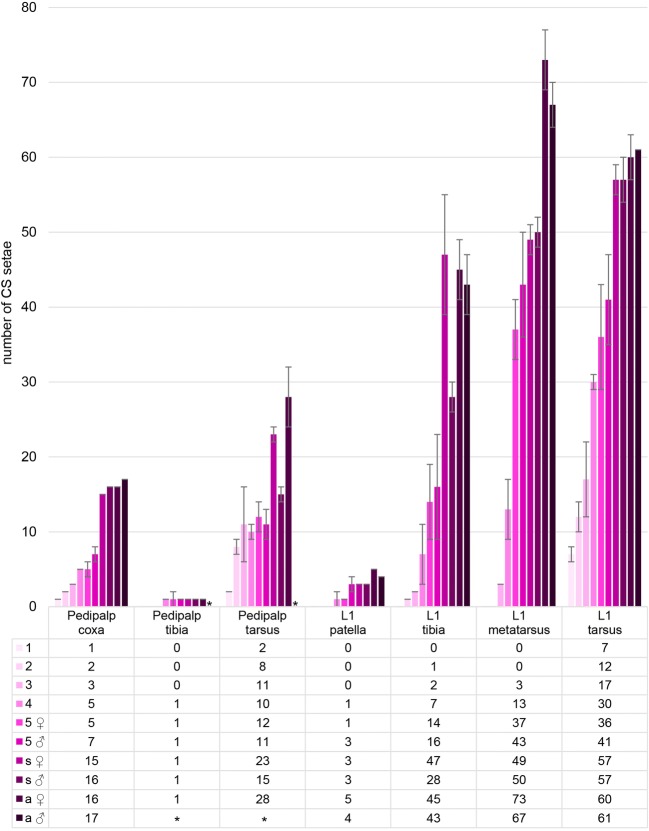
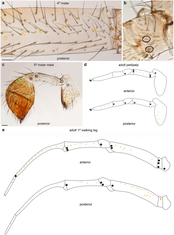
Spiders are equipped with a large number of innervated cuticular specializations, which respond to various sensory stimuli. The physiological function of mechanosensory organs has been analysed in great detail in some model spider species (e.g. Cupiennius salei); however, much less is known about the distribution and function of chemosensory organs. Furthermore, our knowledge on how the sense organ pattern develops on the spider appendages is limited. Here we analyse the development of the pattern and distribution of six different external mechano- and chemosensory organs in all postembryonic stages and in adult male and female spiders of the species Parasteatoda tepidariorum. We show that except for small mechanosensory setae, external sense organs appear in fixed positions on the pedipalps and first walking legs, arranged in longitudinal rows along the proximal-distal axis or in invariable positions relative to morphological landmarks (joints, distal tarsal tip). A comparison to other Entelegynae spiders shows that these features are conserved. We hope that this study lays the foundation for future molecular analysis to address the question how this conserved pattern is generated.
期刊介绍:
Development Genes and Evolution publishes high-quality reports on all aspects of development biology and evolutionary biology. The journal reports on experimental and bioinformatics work at the systemic, cellular and molecular levels in the field of animal and plant systems, covering key aspects of the following topics:
Embryological and genetic analysis of model and non-model organisms
Genes and pattern formation in invertebrates, vertebrates and plants
Axial patterning, embryonic induction and fate maps
Cellular mechanisms of morphogenesis and organogenesis
Stem cells and regeneration
Functional genomics of developmental processes
Developmental diversity and evolution
Evolution of developmentally relevant genes
Phylogeny of animals and plants
Microevolution
Paleontology.

 求助内容:
求助内容: 应助结果提醒方式:


