Paul Spiesecke, Thomas Fischer, Carsten Stephan, Andreas Maxeiner, Bernd Hamm, Markus Lerchbaumer
{"title":"Multiparametric Ultrasound (mpUS) of a Rare Testicular Capillary Hemangioma.","authors":"Paul Spiesecke, Thomas Fischer, Carsten Stephan, Andreas Maxeiner, Bernd Hamm, Markus Lerchbaumer","doi":"10.1155/2019/7568098","DOIUrl":null,"url":null,"abstract":"<p><p>Capillary hemangioma is a rare entity among testicular tumors. We demonstrate the case of an 18-year-old patient with palpatoric and sonographic conspicuous left testicle and negative serum tumor markers (<i>α</i>-fetoprotein, <i>β</i>-human chorionic gonadotropin, and lactate dehydrogenase). Ultrasound (US) imaging represented an isoechogenic lesion with high vascularization in both power Doppler and microflow imaging with central feeding artery. Both strain elastography and shear wave elastography demonstrated a stiff lesion compared to surrounding testicular tissue. While contrast-enhanced ultrasound (CEUS) clearly depicted high vascular load, time intensity curve (TIC) analysis was able to show shorter median transit time, higher peak enhancement, and higher wash-in area under the curve compared to regular testicular tissue. Histopathological examination revealed a lobular constructed and rich vascularized proliferation without cellular atypia and feeder vessels with positive reaction to CD34, CD31, CD99, and Vimentin. Proliferative activity was quantified to 3-5% by Ki-67 index. Two days after surgery, the patient could leave the hospital in subjective wellbeing. While histology remains the gold standard to make a precise diagnosis of capillary hemangiomas due to small case numbers and variety of this benign tumor, the combination of multiparametric US and clinical information may be a promising future tool in preoperative assessment.</p>","PeriodicalId":30326,"journal":{"name":"Case Reports in Radiology","volume":" ","pages":"7568098"},"PeriodicalIF":0.0000,"publicationDate":"2019-12-28","publicationTypes":"Journal Article","fieldsOfStudy":null,"isOpenAccess":false,"openAccessPdf":"https://sci-hub-pdf.com/10.1155/2019/7568098","citationCount":"5","resultStr":null,"platform":"Semanticscholar","paperid":null,"PeriodicalName":"Case Reports in Radiology","FirstCategoryId":"1085","ListUrlMain":"https://doi.org/10.1155/2019/7568098","RegionNum":0,"RegionCategory":null,"ArticlePicture":[],"TitleCN":null,"AbstractTextCN":null,"PMCID":null,"EPubDate":"2019/1/1 0:00:00","PubModel":"eCollection","JCR":"","JCRName":"","Score":null,"Total":0}
引用次数: 5
Abstract
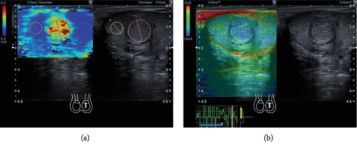
Capillary hemangioma is a rare entity among testicular tumors. We demonstrate the case of an 18-year-old patient with palpatoric and sonographic conspicuous left testicle and negative serum tumor markers (α-fetoprotein, β-human chorionic gonadotropin, and lactate dehydrogenase). Ultrasound (US) imaging represented an isoechogenic lesion with high vascularization in both power Doppler and microflow imaging with central feeding artery. Both strain elastography and shear wave elastography demonstrated a stiff lesion compared to surrounding testicular tissue. While contrast-enhanced ultrasound (CEUS) clearly depicted high vascular load, time intensity curve (TIC) analysis was able to show shorter median transit time, higher peak enhancement, and higher wash-in area under the curve compared to regular testicular tissue. Histopathological examination revealed a lobular constructed and rich vascularized proliferation without cellular atypia and feeder vessels with positive reaction to CD34, CD31, CD99, and Vimentin. Proliferative activity was quantified to 3-5% by Ki-67 index. Two days after surgery, the patient could leave the hospital in subjective wellbeing. While histology remains the gold standard to make a precise diagnosis of capillary hemangiomas due to small case numbers and variety of this benign tumor, the combination of multiparametric US and clinical information may be a promising future tool in preoperative assessment.


 求助内容:
求助内容: 应助结果提醒方式:


