下载PDF
{"title":"A Sensitive and Quantitative mKeima Assay for Mitophagy via FACS","authors":"Chunxin Wang","doi":"10.1002/cpcb.99","DOIUrl":null,"url":null,"abstract":"<p>Mitophagy is a selective autophagy process that specifically removes damaged mitochondria via general autophagy. The two major recessive Parkinson's disease genes PINK1 and Parkin play essential roles in mitophagy initiation, increasing the interest in mitophagy in both basic and translational research over the past 10 years. Initially, mitophagy was measured by the loss of mitochondria either through confocal imaging or immunoblot of mitochondrial proteins such as Tom20 or COXII. Confocal imaging of mitochondria DNA loss via anti-DNA staining is another option. All of these methods, which take considerable effort and labor, are not sensitive enough to detect early stages of mitophagy with unambiguous objectivity. The mKeima assay can be used for both confocal imaging and FACS analysis to provide a thorough picture of mitophagy with a wide dynamic range. The Keima fluorophore has bimodal excitation under neutral and acidic pH conditions. Thus, when Keima is targeted to mitochondria it can accurately reveal the formation of mitolysosomes. Here the author briefly describes the origin and history of mKeima and how it is adapted to measure mitophagy. The author presents detailed protocols for making stable cell lines for optimized mitophagy detection and discuss many parameters that might affect the assay. A troubleshooting section is also provided to discuss possible pitfalls to improve reproducibility and sensitivity of the assay. © 2019 The Authors.</p><p>This article was corrected on 20 August 2022. See the end of the full text for details.</p><p><b>Basic Protocol 1</b>: Making stable lines expressing mito-mKeima and YFP-Parkin</p><p><b>Support Protocol</b>: Retrovirus/lentivirus infection</p><p><b>Basic Protocol 2</b>: FACS analysis of mitophagy</p>","PeriodicalId":40051,"journal":{"name":"Current Protocols in Cell Biology","volume":"86 1","pages":""},"PeriodicalIF":0.0000,"publicationDate":"2019-12-26","publicationTypes":"Journal Article","fieldsOfStudy":null,"isOpenAccess":false,"openAccessPdf":"https://sci-hub-pdf.com/10.1002/cpcb.99","citationCount":"11","resultStr":null,"platform":"Semanticscholar","paperid":null,"PeriodicalName":"Current Protocols in Cell Biology","FirstCategoryId":"1085","ListUrlMain":"https://onlinelibrary.wiley.com/doi/10.1002/cpcb.99","RegionNum":0,"RegionCategory":null,"ArticlePicture":[],"TitleCN":null,"AbstractTextCN":null,"PMCID":null,"EPubDate":"","PubModel":"","JCR":"Q3","JCRName":"Biochemistry, Genetics and Molecular Biology","Score":null,"Total":0}
引用次数: 11
引用
批量引用
Abstract
Mitophagy is a selective autophagy process that specifically removes damaged mitochondria via general autophagy. The two major recessive Parkinson's disease genes PINK1 and Parkin play essential roles in mitophagy initiation, increasing the interest in mitophagy in both basic and translational research over the past 10 years. Initially, mitophagy was measured by the loss of mitochondria either through confocal imaging or immunoblot of mitochondrial proteins such as Tom20 or COXII. Confocal imaging of mitochondria DNA loss via anti-DNA staining is another option. All of these methods, which take considerable effort and labor, are not sensitive enough to detect early stages of mitophagy with unambiguous objectivity. The mKeima assay can be used for both confocal imaging and FACS analysis to provide a thorough picture of mitophagy with a wide dynamic range. The Keima fluorophore has bimodal excitation under neutral and acidic pH conditions. Thus, when Keima is targeted to mitochondria it can accurately reveal the formation of mitolysosomes. Here the author briefly describes the origin and history of mKeima and how it is adapted to measure mitophagy. The author presents detailed protocols for making stable cell lines for optimized mitophagy detection and discuss many parameters that might affect the assay. A troubleshooting section is also provided to discuss possible pitfalls to improve reproducibility and sensitivity of the assay. © 2019 The Authors.
This article was corrected on 20 August 2022. See the end of the full text for details.
Basic Protocol 1 : Making stable lines expressing mito-mKeima and YFP-Parkin
Support Protocol : Retrovirus/lentivirus infection
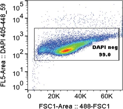
Basic Protocol 2 : FACS analysis of mitophagy
FACS法测定线粒体自噬的灵敏定量mKeima试验
线粒体自噬是一种选择性自噬过程,通过一般自噬特异性地去除受损的线粒体。两个主要的隐性帕金森病基因PINK1和Parkin在线粒体自噬起始中起着至关重要的作用,在过去的10年里,人们对线粒体自噬的基础和转化研究越来越感兴趣。最初,线粒体自噬是通过共聚焦成像或线粒体蛋白(如Tom20或COXII)的免疫印迹来测量线粒体的损失。通过抗DNA染色对线粒体DNA丢失进行共聚焦成像是另一种选择。所有这些方法都需要付出相当大的努力和劳动,它们不够敏感,无法以明确的客观性检测出早期阶段的有丝分裂。mKeima实验可用于共聚焦成像和FACS分析,以提供具有宽动态范围的线粒体自噬的全面图像。Keima荧光团在中性和酸性条件下具有双峰激发。因此,当Keima靶向线粒体时,它可以准确地揭示线粒体溶酶体的形成。在这里,作者简要地描述了mKeima的起源和历史,以及它如何被用来测量有丝分裂。作者提出了制作稳定细胞系的详细方案,以优化有丝分裂检测,并讨论了许多可能影响检测的参数。故障排除部分还提供了讨论可能的陷阱,以提高重现性和灵敏度的测定。©2019作者。本文于2022年8月20日更正。详情见全文末尾。基本方案1:制备表达mito-mKeima和yfp - parkin的稳定细胞系支持方案:逆转录病毒/慢病毒感染基本方案2:有丝分裂的FACS分析
本文章由计算机程序翻译,如有差异,请以英文原文为准。

 求助内容:
求助内容: 应助结果提醒方式:


