Kristin Suetens, Jeroen Swinnen, Linde Stessens, Sofie Van Cauter, Geert Gelin
{"title":"Chordoid Glioma as a Differential Diagnosis of Anterior Third Ventricle Tumours: A Rare Case Report and Five-Year Follow-Up.","authors":"Kristin Suetens, Jeroen Swinnen, Linde Stessens, Sofie Van Cauter, Geert Gelin","doi":"10.1155/2019/3584837","DOIUrl":null,"url":null,"abstract":"<p><p>Chordoid glioma is a rare and relatively recently defined tumour entity. Worldwide there have only been around 90 cases described until now. A chordoid glioma comprises a low-grade suprasellar neuroepithelial neoplasm originating in the anterior part of the third ventricle, with consistent radiological features on MRI. This lesion should be considered as a differential of third ventricle tumours. The patient described in this paper is quite unique in the sense that despite only partial tumour resection was obtained, the residual tumour was not progressive during several years of follow-up. Preoperative recognition of this disease entity is crucial to modify the treatment approach and improve patient outcome.</p>","PeriodicalId":30326,"journal":{"name":"Case Reports in Radiology","volume":" ","pages":"3584837"},"PeriodicalIF":0.0000,"publicationDate":"2019-12-04","publicationTypes":"Journal Article","fieldsOfStudy":null,"isOpenAccess":false,"openAccessPdf":"https://sci-hub-pdf.com/10.1155/2019/3584837","citationCount":"4","resultStr":null,"platform":"Semanticscholar","paperid":null,"PeriodicalName":"Case Reports in Radiology","FirstCategoryId":"1085","ListUrlMain":"https://doi.org/10.1155/2019/3584837","RegionNum":0,"RegionCategory":null,"ArticlePicture":[],"TitleCN":null,"AbstractTextCN":null,"PMCID":null,"EPubDate":"2019/1/1 0:00:00","PubModel":"eCollection","JCR":"","JCRName":"","Score":null,"Total":0}
引用次数: 4
Abstract
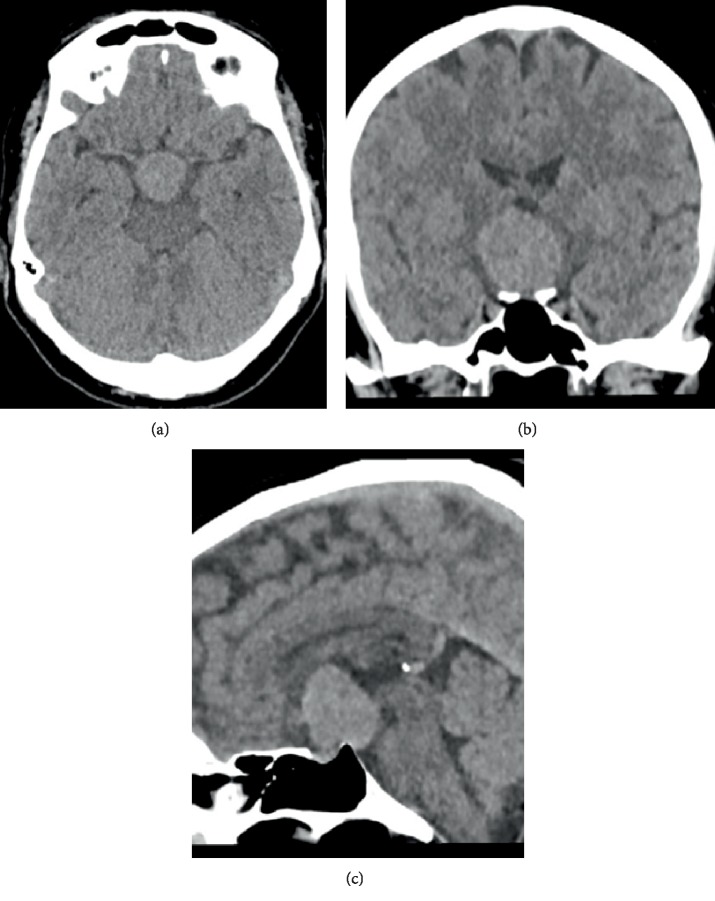
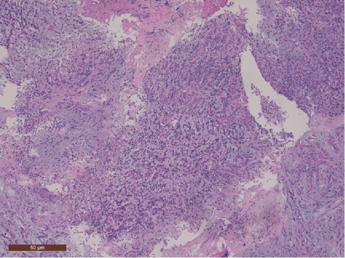
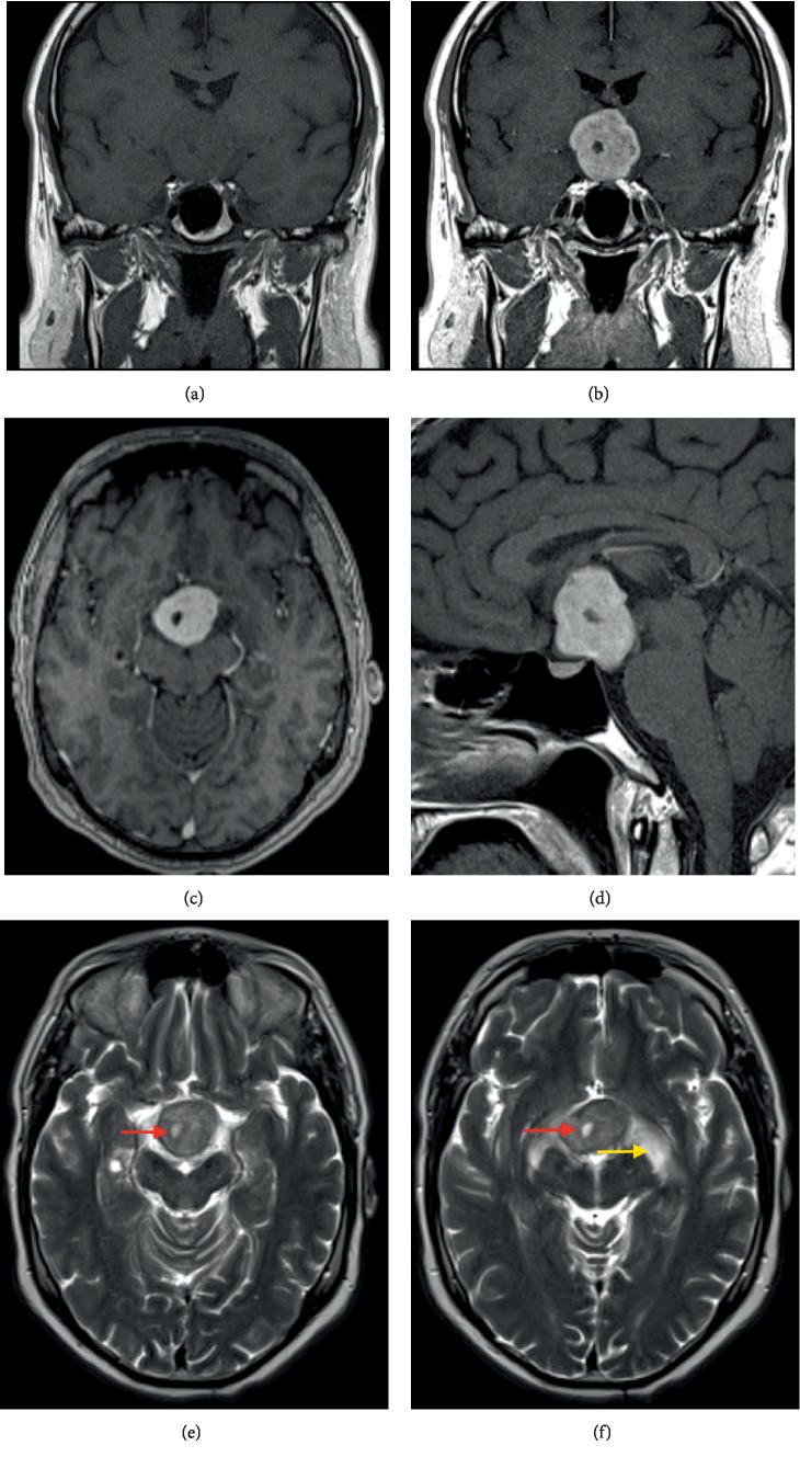
Chordoid glioma is a rare and relatively recently defined tumour entity. Worldwide there have only been around 90 cases described until now. A chordoid glioma comprises a low-grade suprasellar neuroepithelial neoplasm originating in the anterior part of the third ventricle, with consistent radiological features on MRI. This lesion should be considered as a differential of third ventricle tumours. The patient described in this paper is quite unique in the sense that despite only partial tumour resection was obtained, the residual tumour was not progressive during several years of follow-up. Preoperative recognition of this disease entity is crucial to modify the treatment approach and improve patient outcome.

 求助内容:
求助内容: 应助结果提醒方式:


