Fateme Salehi, Mark Landis, Richard Inculet, Daniele Wiseman
{"title":"Case Report of a Rare Cystic Mediastinal Lymphangioma Mimicking Recurrent Pleural Effusion.","authors":"Fateme Salehi, Mark Landis, Richard Inculet, Daniele Wiseman","doi":"10.1155/2019/1301845","DOIUrl":null,"url":null,"abstract":"<p><p>Mediastinal lymphangiomas are rare benign congenital malformations, but complications can occur, including infection, cystic hemorrhage, superior vena cava syndrome, airway compromise, and chylothorax. Radiologically, lymphangiomas are well-defined masses, with low attenuation ranging from simple to complex fluid and fat. They often encase adjacent mediastinal structures. We present a case of mediastinal lymphangioma in a young female, who presented with recurrent complex pleural effusions, initially thought to represent an empyema and/or necrotic mass. Despite surgical chest tube and interventional radiology drainage, fluid reaccumulated. Upon further review, the interventional and thoracic radiologist concurred that the complex collection was in fact predominantly extra pleural in location. The patient underwent partial resection after it was discovered intraoperatively that the extra pleural cystic mass was contiguous with and extended deeply into the mediastinum. Histopathology confirmed the diagnosis of lymphangioma.</p>","PeriodicalId":30326,"journal":{"name":"Case Reports in Radiology","volume":null,"pages":null},"PeriodicalIF":0.0000,"publicationDate":"2019-05-26","publicationTypes":"Journal Article","fieldsOfStudy":null,"isOpenAccess":false,"openAccessPdf":"https://sci-hub-pdf.com/10.1155/2019/1301845","citationCount":"2","resultStr":null,"platform":"Semanticscholar","paperid":null,"PeriodicalName":"Case Reports in Radiology","FirstCategoryId":"1085","ListUrlMain":"https://doi.org/10.1155/2019/1301845","RegionNum":0,"RegionCategory":null,"ArticlePicture":[],"TitleCN":null,"AbstractTextCN":null,"PMCID":null,"EPubDate":"2019/1/1 0:00:00","PubModel":"eCollection","JCR":"","JCRName":"","Score":null,"Total":0}
引用次数: 2
Abstract
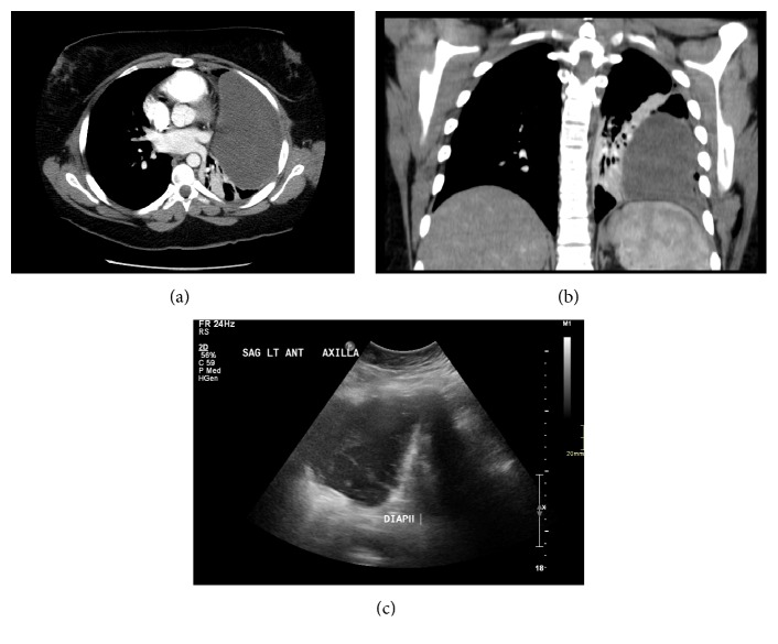
Mediastinal lymphangiomas are rare benign congenital malformations, but complications can occur, including infection, cystic hemorrhage, superior vena cava syndrome, airway compromise, and chylothorax. Radiologically, lymphangiomas are well-defined masses, with low attenuation ranging from simple to complex fluid and fat. They often encase adjacent mediastinal structures. We present a case of mediastinal lymphangioma in a young female, who presented with recurrent complex pleural effusions, initially thought to represent an empyema and/or necrotic mass. Despite surgical chest tube and interventional radiology drainage, fluid reaccumulated. Upon further review, the interventional and thoracic radiologist concurred that the complex collection was in fact predominantly extra pleural in location. The patient underwent partial resection after it was discovered intraoperatively that the extra pleural cystic mass was contiguous with and extended deeply into the mediastinum. Histopathology confirmed the diagnosis of lymphangioma.

 求助内容:
求助内容: 应助结果提醒方式:


