Thomas Kotsis, Panagitsa Christoforou, Constantinos Nastos
{"title":"Carotid Paraganglioma in Adolescence-Clinical Picture-Surgical Technique and Review of the Literature.","authors":"Thomas Kotsis, Panagitsa Christoforou, Constantinos Nastos","doi":"10.1155/2019/6182783","DOIUrl":null,"url":null,"abstract":"<p><p>Paraganglia are clusters of cells originating from the neural crest with histological and cytochemical characteristics of neuroendocrine cells. Paragangliomas of the head and neck represent less than 0.5% of all head and neck tumors and they usually occur between the ages of 40 and 50. Paragangliomas in childhood and in adolescence are extremely rare; only 23 case reports have been reported in the recent literature. In childhood, the estimation of malignant potential is 3-10%; therefore, early diagnosis and treatment of carotid body paragangliomas are mandatory. However, due to the rarity of these lesions in young patients, they are often not included in the differential diagnosis of solid masses in the neck area, a fact that may lead to misdiagnosis or delay in treatment. We present, herein, two extremely rare cases of patients in adolescence who were diagnosed with a carotid body paraganglioma and were treated surgically in our unit. One of the patients was diagnosed and treated at the age of 15 years while the other had a long-standing tumor in the neck that was followed up by a general surgery outpatient service as a branchial cleft cyst at the age of 15 years and was eventually treated surgically 8 years later. Carotid body tumor was not considered in the initial differential diagnosis because of its rarity at this age range.</p>","PeriodicalId":9632,"journal":{"name":"Case Reports in Vascular Medicine","volume":"2019 ","pages":"6182783"},"PeriodicalIF":0.0000,"publicationDate":"2019-03-10","publicationTypes":"Journal Article","fieldsOfStudy":null,"isOpenAccess":false,"openAccessPdf":"https://sci-hub-pdf.com/10.1155/2019/6182783","citationCount":"2","resultStr":null,"platform":"Semanticscholar","paperid":null,"PeriodicalName":"Case Reports in Vascular Medicine","FirstCategoryId":"1085","ListUrlMain":"https://doi.org/10.1155/2019/6182783","RegionNum":0,"RegionCategory":null,"ArticlePicture":[],"TitleCN":null,"AbstractTextCN":null,"PMCID":null,"EPubDate":"2019/1/1 0:00:00","PubModel":"eCollection","JCR":"","JCRName":"","Score":null,"Total":0}
引用次数: 2
Abstract
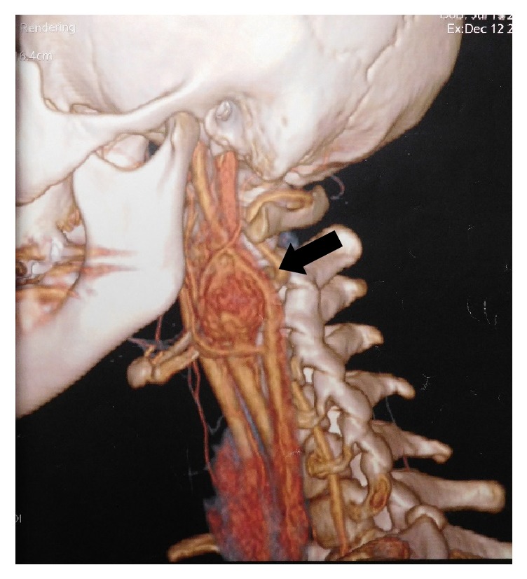
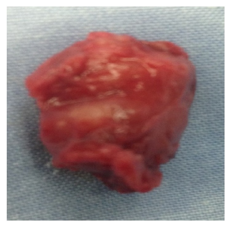
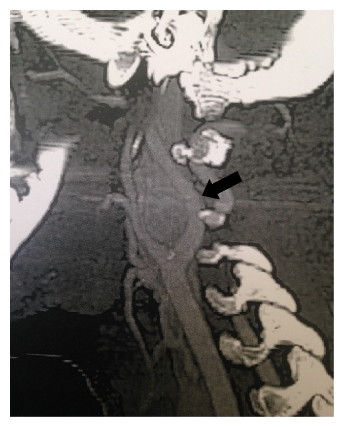
Paraganglia are clusters of cells originating from the neural crest with histological and cytochemical characteristics of neuroendocrine cells. Paragangliomas of the head and neck represent less than 0.5% of all head and neck tumors and they usually occur between the ages of 40 and 50. Paragangliomas in childhood and in adolescence are extremely rare; only 23 case reports have been reported in the recent literature. In childhood, the estimation of malignant potential is 3-10%; therefore, early diagnosis and treatment of carotid body paragangliomas are mandatory. However, due to the rarity of these lesions in young patients, they are often not included in the differential diagnosis of solid masses in the neck area, a fact that may lead to misdiagnosis or delay in treatment. We present, herein, two extremely rare cases of patients in adolescence who were diagnosed with a carotid body paraganglioma and were treated surgically in our unit. One of the patients was diagnosed and treated at the age of 15 years while the other had a long-standing tumor in the neck that was followed up by a general surgery outpatient service as a branchial cleft cyst at the age of 15 years and was eventually treated surgically 8 years later. Carotid body tumor was not considered in the initial differential diagnosis because of its rarity at this age range.

 求助内容:
求助内容: 应助结果提醒方式:


