Probable sporadic Creutzfeldt–Jakob disease mimicking focal epilepsy
引用次数: 8
Abstract
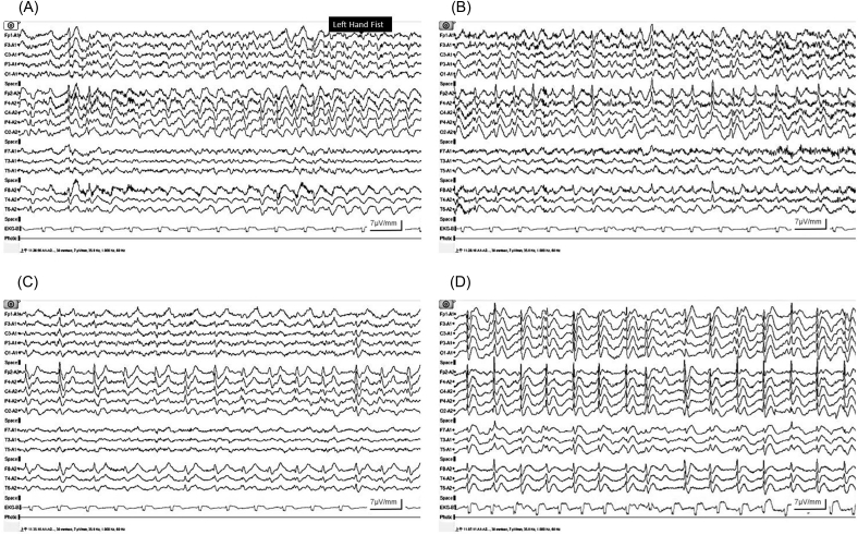
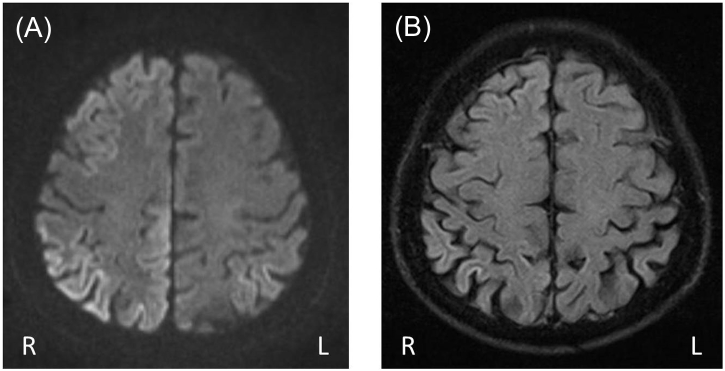
Creutzfeldt–Jakob disease (CJD) presents with seizures as an early symptom in only approximately 3% of cases. These seizures often present as nonconvulsive status epilepticus (NCSE) or epilepsia partialis continua (EPC). Here, we describe a case of probable sporadic CJD (sCJD) in an 83-year-old man whose manifest an unusual presentation of left-hand tonic seizures without evolution to EPC, as well as brain MRI findings interpreted as peri-ictal changes, which led to an initial misdiagnosis of focal epilepsy.
可能是散发的克雅氏病,类似局灶性癫痫
克雅氏病(CJD)的早期症状表现为癫痫发作,只有约3%的病例。这些癫痫发作通常表现为非惊厥性癫痫持续状态(NCSE)或部分持续性癫痫(EPC)。在这里,我们描述了一例可能是散发CJD (sCJD)的83岁男性,其表现为不寻常的左强张性癫痫发作,没有演变为EPC,以及脑MRI结果解释为周周改变,导致最初误诊为局灶性癫痫。
本文章由计算机程序翻译,如有差异,请以英文原文为准。
求助全文
约1分钟内获得全文
求助全文

 求助内容:
求助内容: 应助结果提醒方式:


