Effects of Electroacupuncture on Expression of D1 Receptor (D1R), Phosphorylation of Extracellular-Regulated Protein Kinase 1/2 (p-ERK1/2), and c-Fos in the Insular Cortex of Ketamine-Addicted Rats.
{"title":"Effects of Electroacupuncture on Expression of D1 Receptor (D1R), Phosphorylation of Extracellular-Regulated Protein Kinase 1/2 (p-ERK1/2), and c-Fos in the Insular Cortex of Ketamine-Addicted Rats.","authors":"Feng Wu, Jian Ding, Huai-Bin Li, Hua-Chun Miao, Rui Bao, Shan Yang","doi":"10.12659/MSMBR.913285","DOIUrl":null,"url":null,"abstract":"<p><p>BACKGROUND The aim of this study was to investigate the effects of electroacupuncture (EA) on expression of the D1 receptor (D1R), phosphorylation of extracellular-regulated protein kinase 1/2 (p-ERK1/2) and c-Fos in the insular cortex (IC) of ketamine-addicted rats. MATERIAL AND METHODS Sprague-Dawley rats were randomly divided into 7 groups: the normal group, the normal saline (NS) group, the ketamine (Ket) group, the U0126+Ket group, the SCH23390+Ket group, the Ket+acupoints EA (EA1) group, and the Ket+ non-acupoints EA (EA2) group. We used immunohistochemistry to detect the expression of D1R, p-ERK1/2, and c-Fos. We also used Nissl staining techniques to study the morphology of IC neurons. RESULTS Our study demonstrated that the ketamine group had sparsely distributed neurons, large intracellular vacuoles, nuclei shift, and unclear nucleolus. The number of Nissl-positive (neuronal) cells in the ketamine group were decreased than in the normal group. Our results also indicated that there was significantly lower expression of D1R, p-ERK1/2, and c-Fos in the IC of the U0126+Ket group, SCH23390+Ket group, and Ket+EA1 group as compared with that of the Ket group. CONCLUSIONS Ketamine addiction induces c-Fos overexpression in the IC by increasing the expression of D1R and p-ERK1/2. Acupoints EA downregulate D1R and p-ERK1/2 by reducing the overexpression of c-Fos.</p>","PeriodicalId":18491,"journal":{"name":"Medical Science Monitor Basic Research","volume":"25 ","pages":"26-32"},"PeriodicalIF":2.0000,"publicationDate":"2019-01-31","publicationTypes":"Journal Article","fieldsOfStudy":null,"isOpenAccess":false,"openAccessPdf":"https://ftp.ncbi.nlm.nih.gov/pub/pmc/oa_pdf/39/ef/medscimonitbasicres-25-26.PMC6369650.pdf","citationCount":"3","resultStr":null,"platform":"Semanticscholar","paperid":null,"PeriodicalName":"Medical Science Monitor Basic Research","FirstCategoryId":"1085","ListUrlMain":"https://doi.org/10.12659/MSMBR.913285","RegionNum":0,"RegionCategory":null,"ArticlePicture":[],"TitleCN":null,"AbstractTextCN":null,"PMCID":null,"EPubDate":"","PubModel":"","JCR":"Q3","JCRName":"MEDICINE, RESEARCH & EXPERIMENTAL","Score":null,"Total":0}
引用次数: 3
Abstract
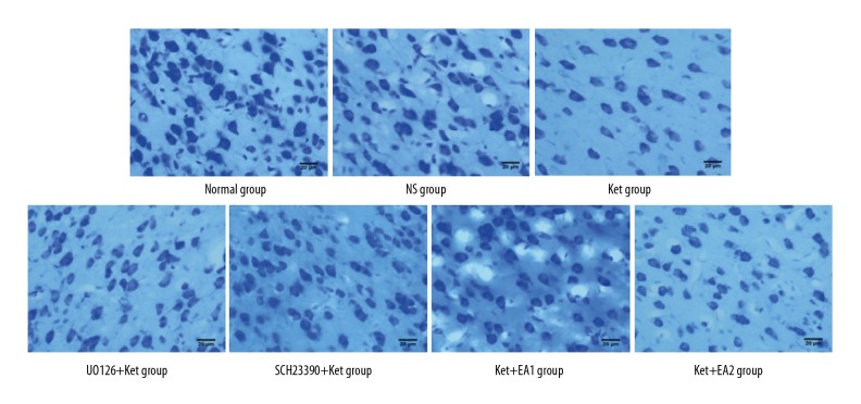
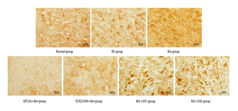
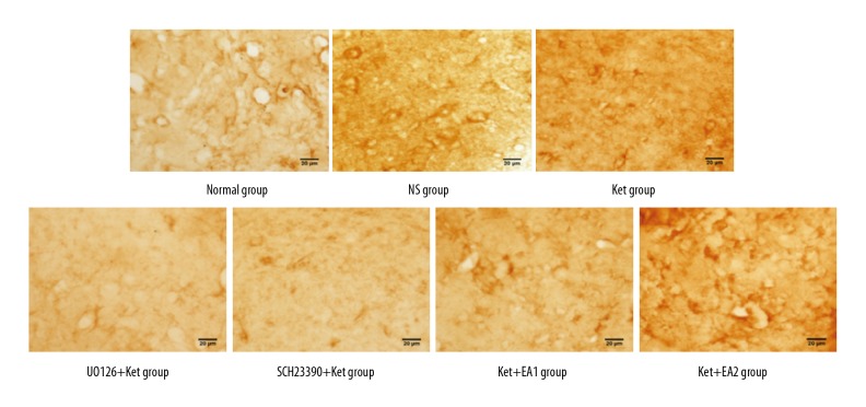
BACKGROUND The aim of this study was to investigate the effects of electroacupuncture (EA) on expression of the D1 receptor (D1R), phosphorylation of extracellular-regulated protein kinase 1/2 (p-ERK1/2) and c-Fos in the insular cortex (IC) of ketamine-addicted rats. MATERIAL AND METHODS Sprague-Dawley rats were randomly divided into 7 groups: the normal group, the normal saline (NS) group, the ketamine (Ket) group, the U0126+Ket group, the SCH23390+Ket group, the Ket+acupoints EA (EA1) group, and the Ket+ non-acupoints EA (EA2) group. We used immunohistochemistry to detect the expression of D1R, p-ERK1/2, and c-Fos. We also used Nissl staining techniques to study the morphology of IC neurons. RESULTS Our study demonstrated that the ketamine group had sparsely distributed neurons, large intracellular vacuoles, nuclei shift, and unclear nucleolus. The number of Nissl-positive (neuronal) cells in the ketamine group were decreased than in the normal group. Our results also indicated that there was significantly lower expression of D1R, p-ERK1/2, and c-Fos in the IC of the U0126+Ket group, SCH23390+Ket group, and Ket+EA1 group as compared with that of the Ket group. CONCLUSIONS Ketamine addiction induces c-Fos overexpression in the IC by increasing the expression of D1R and p-ERK1/2. Acupoints EA downregulate D1R and p-ERK1/2 by reducing the overexpression of c-Fos.

 求助内容:
求助内容: 应助结果提醒方式:


