Nagaraja Chappidi, Giuseppe De Gregorio, Stefano Ferrari
{"title":"Replication stress-induced Exo1 phosphorylation is mediated by Rad53/Pph3 and Exo1 nuclear localization is controlled by 14-3-3 proteins.","authors":"Nagaraja Chappidi, Giuseppe De Gregorio, Stefano Ferrari","doi":"10.1186/s13008-018-0044-2","DOIUrl":null,"url":null,"abstract":"<p><strong>Background: </strong>Mechanisms controlling DNA resection at sites of damage and affecting genome stability have been the subject of deep investigation, though their complexity is not yet fully understood. Specifically, the regulatory role of post-translational modifications in the localization, stability and function of DNA repair proteins is an important aspect of such complexity.</p><p><strong>Results: </strong>Here, we took advantage of the superior resolution of phosphorylated proteins provided by Phos-Tag technology to study pathways controlling the reversible phosphorylation of yeast Exo1, an exonuclease involved in a number of DNA repair pathways. We report that Rad53, a checkpoint kinase downstream of Mec1, is responsible for Exo1 phosphorylation in response to DNA replication stress and we demonstrate a role for the type-2A protein phosphatase Pph3 in the dephosphorylation of both Rad53 and Exo1 during checkpoint recovery. Fluorescence microscopy studies showed that Rad53-dependent phosphorylation is not required for the recruitment or the release of Exo1 from the nucleus, whereas 14-3-3 proteins are necessary for Exo1 nuclear translocation.</p><p><strong>Conclusions: </strong>By shedding light on the mechanism of Exo1 control, these data underscore the importance of post-translational modifications and protein interactions in the regulation of DNA end resection.</p>","PeriodicalId":49263,"journal":{"name":"Cell Division","volume":"14 ","pages":"1"},"PeriodicalIF":2.8000,"publicationDate":"2019-01-04","publicationTypes":"Journal Article","fieldsOfStudy":null,"isOpenAccess":false,"openAccessPdf":"https://sci-hub-pdf.com/10.1186/s13008-018-0044-2","citationCount":"7","resultStr":null,"platform":"Semanticscholar","paperid":null,"PeriodicalName":"Cell Division","FirstCategoryId":"99","ListUrlMain":"https://doi.org/10.1186/s13008-018-0044-2","RegionNum":4,"RegionCategory":"生物学","ArticlePicture":[],"TitleCN":null,"AbstractTextCN":null,"PMCID":null,"EPubDate":"2019/1/1 0:00:00","PubModel":"eCollection","JCR":"Q3","JCRName":"CELL BIOLOGY","Score":null,"Total":0}
引用次数: 7
Abstract
Background: Mechanisms controlling DNA resection at sites of damage and affecting genome stability have been the subject of deep investigation, though their complexity is not yet fully understood. Specifically, the regulatory role of post-translational modifications in the localization, stability and function of DNA repair proteins is an important aspect of such complexity.
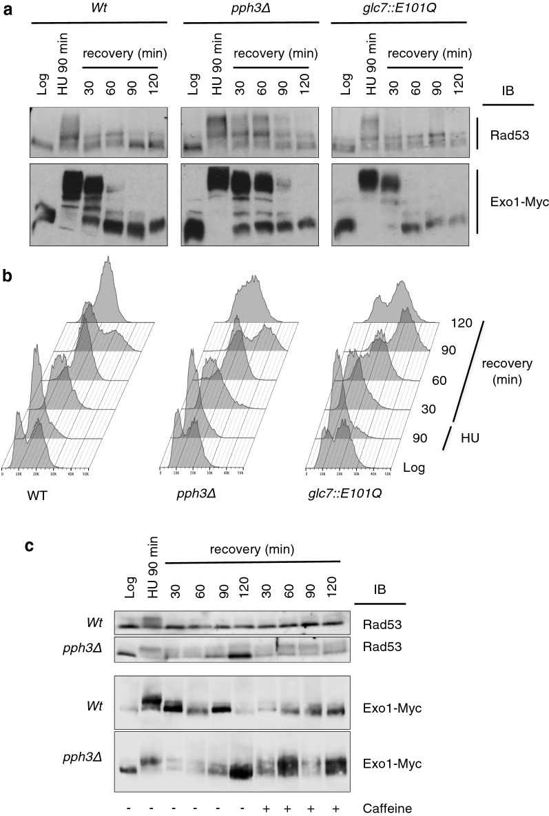
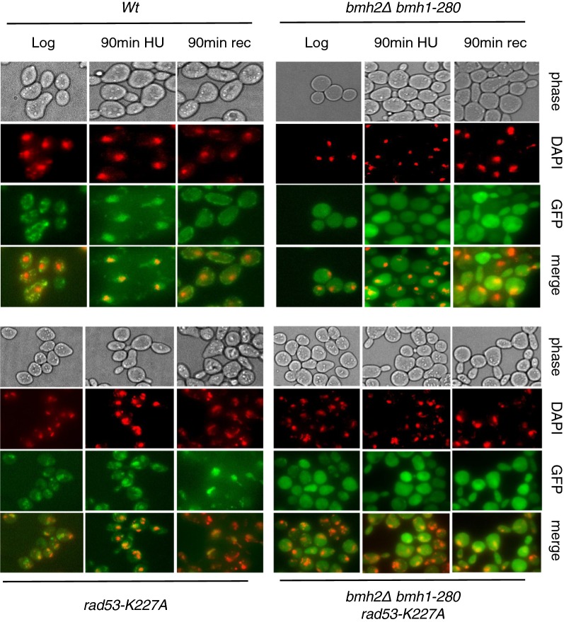
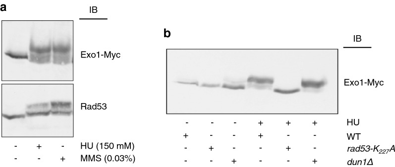
Results: Here, we took advantage of the superior resolution of phosphorylated proteins provided by Phos-Tag technology to study pathways controlling the reversible phosphorylation of yeast Exo1, an exonuclease involved in a number of DNA repair pathways. We report that Rad53, a checkpoint kinase downstream of Mec1, is responsible for Exo1 phosphorylation in response to DNA replication stress and we demonstrate a role for the type-2A protein phosphatase Pph3 in the dephosphorylation of both Rad53 and Exo1 during checkpoint recovery. Fluorescence microscopy studies showed that Rad53-dependent phosphorylation is not required for the recruitment or the release of Exo1 from the nucleus, whereas 14-3-3 proteins are necessary for Exo1 nuclear translocation.
Conclusions: By shedding light on the mechanism of Exo1 control, these data underscore the importance of post-translational modifications and protein interactions in the regulation of DNA end resection.
期刊介绍:
Cell Division is an open access, peer-reviewed journal that encompasses all the molecular aspects of cell cycle control and cancer, cell growth, proliferation, survival, differentiation, signalling, gene transcription, protein synthesis, genome integrity, chromosome stability, centrosome duplication, DNA damage and DNA repair.
Cell Division provides an online forum for the cell-cycle community that aims to publish articles on all exciting aspects of cell-cycle research and to bridge the gap between models of cell cycle regulation, development, and cancer biology. This forum is driven by specialized and timely research articles, reviews and commentaries focused on this fast moving field, providing an invaluable tool for cell-cycle biologists.
Cell Division publishes articles in areas which includes, but not limited to:
DNA replication, cell fate decisions, cell cycle & development
Cell proliferation, mitosis, spindle assembly checkpoint, ubiquitin mediated degradation
DNA damage & repair
Apoptosis & cell death
 求助内容:
求助内容: 应助结果提醒方式:


