Steven M Presciutti, Philip K Louie, Jannat M Khan, Bryce A Basques, Comron Saifi, Christopher J Dewald, Dino Samartzis, Howard S An
{"title":"Sagittal spinopelvic malalignment in degenerative scoliosis patients: isolated correction of symptomatic levels and clinical decision-making.","authors":"Steven M Presciutti, Philip K Louie, Jannat M Khan, Bryce A Basques, Comron Saifi, Christopher J Dewald, Dino Samartzis, Howard S An","doi":"10.1186/s13013-018-0174-y","DOIUrl":null,"url":null,"abstract":"<p><strong>Background: </strong>This study aims to determine if (1) loss of lumbar lordosis (LL), often associated with degenerative scoliosis (DS), is structural or rather largely due to positional factors secondary to spinal stenosis; (2) only addressing the symptomatic levels with a decompression and posterolateral fusion in carefully selected patients will result in improvement of sagittal malalignment; and (3) degree of sagittal plane correction achieved with such a local fusion could be predicted by routine pre-operative imaging.</p><p><strong>Methods: </strong>A retrospective study design with prospectively collected imaging data of a consecutive series of surgically treated DS patients who underwent decompression and instrumented fusion at only symptomatic levels was performed. Pre- and post-operative plain radiographs and pre-operative magnetic resonance imaging (MRIs) of the spinopelvic region were analyzed. LL, pelvic incidence (PI), pelvic tilt (PT), and sacral slope (SS) were assessed in all patients. As a requirement for the surgical strategy, all patients presented with a pre-operative PI-LL mismatch greater than 10<sup>°</sup>. Post-operative complications were assessed.</p><p><strong>Results: </strong>Pre-operative MRIs and lumbar extension radiographs revealed a mean LL of 42<sup>°</sup> (range 10-66<sup>°</sup>) and 48<sup>°</sup> (range 20-74<sup>°</sup>), respectively, in 68 patients (mean follow-up 29 months). LL post-operatively was corrected to a mean PI-LL of 10<sup>°</sup>. Of patients who achieved PI-LL mismatch within 10<sup>o</sup> on their pre-operative extension lateral lumbar radiographs, 62.5% were able to maintain a PI-LL mismatch within 10<sup>°</sup> on their initial post-operative films. Only 37.5% were not able to achieve that mismatch on extension radiographs (<i>p</i> = 0.001, OR = 9.58). Similarly, 54.2% were able to achieve a PI-LL < 10<sup>°</sup> on initial post-operative radiographs, when pre-operative MRI revealed a PI-LL mismatch within 10<sup>°</sup>. In contrast, only 20.5% achieved that goal post-operatively if their mismatch was greater than 10<sup>o</sup> on their MRI (<i>p</i> = 0.003, OR = 4.25).</p><p><strong>Conclusion: </strong>With a decompression and instrumented fusion of only the symptomatic levels in symptomatic DS patients, we were able to achieve a PI-LL mismatch to within 10<sup>°</sup>. The loss of LL observed pre-operatively may be largely positional rather than structural. The amount of LL correction observed immediately after surgery can be predicted from pre-operative lumbar extension radiographs and supine sagittal MRI.</p>","PeriodicalId":21573,"journal":{"name":"Scoliosis and Spinal Disorders","volume":"13 ","pages":"28"},"PeriodicalIF":0.0000,"publicationDate":"2018-12-27","publicationTypes":"Journal Article","fieldsOfStudy":null,"isOpenAccess":false,"openAccessPdf":"https://sci-hub-pdf.com/10.1186/s13013-018-0174-y","citationCount":"5","resultStr":null,"platform":"Semanticscholar","paperid":null,"PeriodicalName":"Scoliosis and Spinal Disorders","FirstCategoryId":"1085","ListUrlMain":"https://doi.org/10.1186/s13013-018-0174-y","RegionNum":0,"RegionCategory":null,"ArticlePicture":[],"TitleCN":null,"AbstractTextCN":null,"PMCID":null,"EPubDate":"2018/1/1 0:00:00","PubModel":"eCollection","JCR":"Q1","JCRName":"Medicine","Score":null,"Total":0}
引用次数: 5
Abstract
Background: This study aims to determine if (1) loss of lumbar lordosis (LL), often associated with degenerative scoliosis (DS), is structural or rather largely due to positional factors secondary to spinal stenosis; (2) only addressing the symptomatic levels with a decompression and posterolateral fusion in carefully selected patients will result in improvement of sagittal malalignment; and (3) degree of sagittal plane correction achieved with such a local fusion could be predicted by routine pre-operative imaging.
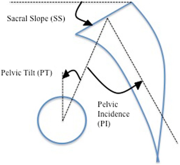
Methods: A retrospective study design with prospectively collected imaging data of a consecutive series of surgically treated DS patients who underwent decompression and instrumented fusion at only symptomatic levels was performed. Pre- and post-operative plain radiographs and pre-operative magnetic resonance imaging (MRIs) of the spinopelvic region were analyzed. LL, pelvic incidence (PI), pelvic tilt (PT), and sacral slope (SS) were assessed in all patients. As a requirement for the surgical strategy, all patients presented with a pre-operative PI-LL mismatch greater than 10°. Post-operative complications were assessed.
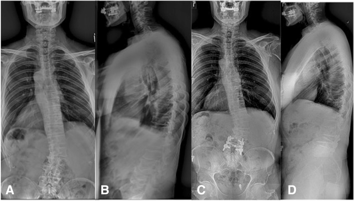
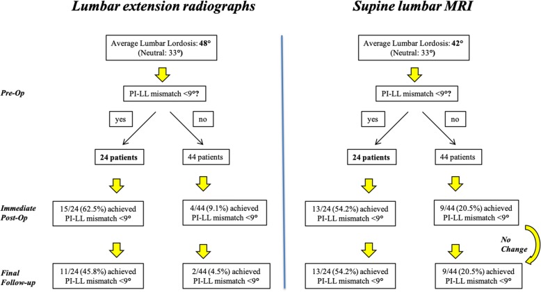
Results: Pre-operative MRIs and lumbar extension radiographs revealed a mean LL of 42° (range 10-66°) and 48° (range 20-74°), respectively, in 68 patients (mean follow-up 29 months). LL post-operatively was corrected to a mean PI-LL of 10°. Of patients who achieved PI-LL mismatch within 10o on their pre-operative extension lateral lumbar radiographs, 62.5% were able to maintain a PI-LL mismatch within 10° on their initial post-operative films. Only 37.5% were not able to achieve that mismatch on extension radiographs (p = 0.001, OR = 9.58). Similarly, 54.2% were able to achieve a PI-LL < 10° on initial post-operative radiographs, when pre-operative MRI revealed a PI-LL mismatch within 10°. In contrast, only 20.5% achieved that goal post-operatively if their mismatch was greater than 10o on their MRI (p = 0.003, OR = 4.25).
Conclusion: With a decompression and instrumented fusion of only the symptomatic levels in symptomatic DS patients, we were able to achieve a PI-LL mismatch to within 10°. The loss of LL observed pre-operatively may be largely positional rather than structural. The amount of LL correction observed immediately after surgery can be predicted from pre-operative lumbar extension radiographs and supine sagittal MRI.
期刊介绍:
Cessation.Scoliosis and Spinal Disorders is an open access, multidisciplinary journal that encompasses all aspects of research on prevention, diagnosis, treatment, outcomes and cost-analyses of conservative and surgical management of all spinal deformities and disorders. Both clinical and basic science reports form the cornerstone of the journal in its endeavour to provide original, primary studies as well as narrative/systematic reviews and meta-analyses to the academic community and beyond. Scoliosis and Spinal Disorders aims to provide an integrated and balanced view of cutting-edge spine research to further enhance effective collaboration among clinical spine specialists and scientists, and to ultimately improve patient outcomes based on an evidence-based spine care approach.

 求助内容:
求助内容: 应助结果提醒方式:


