Florian Hentschel, Anna Franziska Jansen, Marlis Günther, Roland Pauli, Stefan Lüth
{"title":"Eosinophil Counts in Mucosal Biopsies of the Ileum and Colon: Interobserver Variance Affects Diagnostic Accuracy.","authors":"Florian Hentschel, Anna Franziska Jansen, Marlis Günther, Roland Pauli, Stefan Lüth","doi":"10.1155/2018/2638258","DOIUrl":null,"url":null,"abstract":"<p><p>Primary eosinophilic gastroenteritis and colitis (EGE) is a rare entity with unspecific clinical and endoscopic findings. Validated histopathologic criteria for confirming the diagnosis are lacking, because numeric values for normal or elevated concentrations of eosinophils in mucosal biopsies are varying between observers. To quantify this interobserver variance, we had the same set of 30 slides of eosinophilic-rich mucosal biopsies from the ileum and colon systematically reviewed by a panel of six independent pathologists, each with more than a ten-year experience in the field. Using a highly standardized biopsy and slide preparation protocol, we ruled out any influence by the preparation, the patient, the endoscopist, the endoscopes and calipers used, the sampling site, the fixation and staining method, and the microscopic field sizes. Still, all numeric results differed between pathologists up to a factor greater than 30. Calculated positive or negative diagnosis of EGE differed up to a factor greater than 8. A theoretical incidence for EGE calculated from these numbers differed by a factor greater than 1500. We conclude that eosinophil counts in mucosal biopsies from the lower gastrointestinal tract are subject to a very high interobserver variance. Until further research provides objective and validated methods for standardization, all epidemiologic numbers derived from histopathologic findings may have to be questioned. When diagnosing individual patients with EGE, overall morphologic picture together with clinical and endoscopic findings is more important than numeric eosinophil count.</p>","PeriodicalId":89212,"journal":{"name":"Pathology research international","volume":"2018 ","pages":"2638258"},"PeriodicalIF":0.0000,"publicationDate":"2018-11-04","publicationTypes":"Journal Article","fieldsOfStudy":null,"isOpenAccess":false,"openAccessPdf":"https://www.ncbi.nlm.nih.gov/pmc/articles/PMC6241360/pdf/","citationCount":"7","resultStr":null,"platform":"Semanticscholar","paperid":null,"PeriodicalName":"Pathology research international","FirstCategoryId":"1085","ListUrlMain":"https://doi.org/10.1155/2018/2638258","RegionNum":0,"RegionCategory":null,"ArticlePicture":[],"TitleCN":null,"AbstractTextCN":null,"PMCID":null,"EPubDate":"2018/1/1 0:00:00","PubModel":"eCollection","JCR":"","JCRName":"","Score":null,"Total":0}
引用次数: 7
Abstract
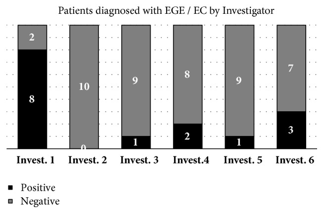
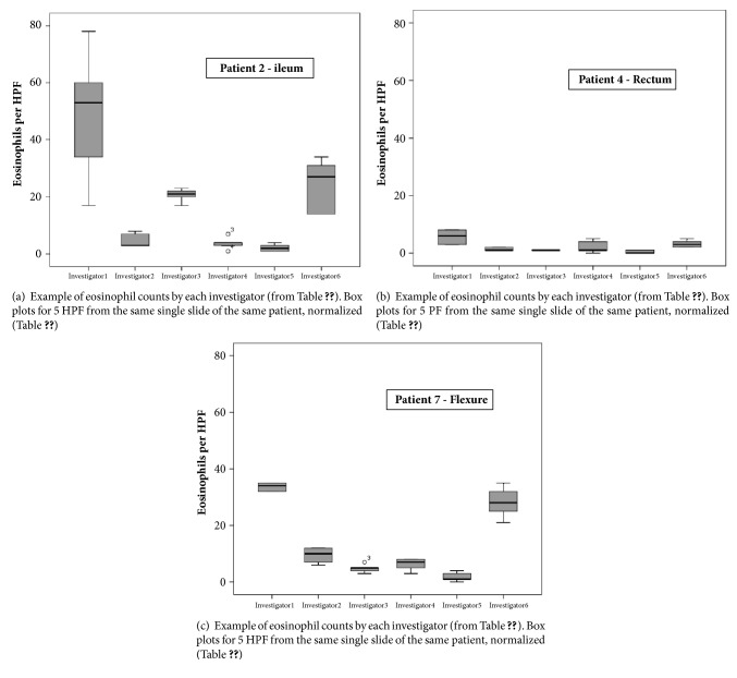
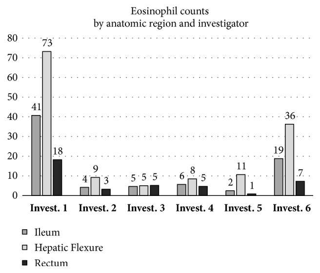
Primary eosinophilic gastroenteritis and colitis (EGE) is a rare entity with unspecific clinical and endoscopic findings. Validated histopathologic criteria for confirming the diagnosis are lacking, because numeric values for normal or elevated concentrations of eosinophils in mucosal biopsies are varying between observers. To quantify this interobserver variance, we had the same set of 30 slides of eosinophilic-rich mucosal biopsies from the ileum and colon systematically reviewed by a panel of six independent pathologists, each with more than a ten-year experience in the field. Using a highly standardized biopsy and slide preparation protocol, we ruled out any influence by the preparation, the patient, the endoscopist, the endoscopes and calipers used, the sampling site, the fixation and staining method, and the microscopic field sizes. Still, all numeric results differed between pathologists up to a factor greater than 30. Calculated positive or negative diagnosis of EGE differed up to a factor greater than 8. A theoretical incidence for EGE calculated from these numbers differed by a factor greater than 1500. We conclude that eosinophil counts in mucosal biopsies from the lower gastrointestinal tract are subject to a very high interobserver variance. Until further research provides objective and validated methods for standardization, all epidemiologic numbers derived from histopathologic findings may have to be questioned. When diagnosing individual patients with EGE, overall morphologic picture together with clinical and endoscopic findings is more important than numeric eosinophil count.

 求助内容:
求助内容: 应助结果提醒方式:


