Quenum Coffi, Sonou Arnaud, Gouthon Polycarpe, Ahissou Hyacinthe, Messan Folly, Nouatin Basile, Hounkponou Murielle, Houénassi D Martin
{"title":"Changes in Echocardiographic Parameters among Beninese Soccer Referees during the Division 1 Championship in 2016.","authors":"Quenum Coffi, Sonou Arnaud, Gouthon Polycarpe, Ahissou Hyacinthe, Messan Folly, Nouatin Basile, Hounkponou Murielle, Houénassi D Martin","doi":"10.1155/2018/6024574","DOIUrl":null,"url":null,"abstract":"<p><strong>Introduction: </strong>The goal of this study was to describe the echocardiographic parameters of soccer referees and to examine the changes in these parameters after a period of intensive physical exercise.</p><p><strong>Methods and patients: </strong>We conducted a prospective study that included Beninese soccer referees. The study of the geometry and function of the left ventricle (LV) was made at the beginning and end of the national Division 1 championship, which was held during the course of 10 weeks.</p><p><strong>Results: </strong>There were 37 referees included in this study; 20 at the national level (G1: 27.8 ± 6.6 years) and 17 at the international level (G2: 32.1 ± 6.4 years). Dimensions of the LV were normal for all the referees. At the beginning of the championship, 51.3% of the referees had a normal LV geometry, 37.8% had concentric remodelling, 2.7% had concentric hypertrophy, and 8.1% had eccentric hypertrophy. In the group of referees with normal LV geometry, a modification in concentric remodelling at the end of the championship was seen in 30% of the referees in G1, 33.3% of the referees in G2, and 31.6% of the whole sample. In the group of subjects who presented concentric LV remodelling, a modification in the normal geometry was observed in 37.5% of those in G1, in 0% of those in G2, and in 21.4% of the whole sample. The cases of LV hypertrophy showed no change regardless of the group considered. An LV ejection fraction of more than 50% and an E/E' ratio less than 8 were found in all referees.</p><p><strong>Conclusion: </strong>All the referees studied had normal cardiac morphology and function. The intensity of the physical load was insufficient to impact this morphology.</p>","PeriodicalId":30574,"journal":{"name":"Journal of Sports Medicine","volume":"2018 ","pages":"6024574"},"PeriodicalIF":0.0000,"publicationDate":"2018-11-07","publicationTypes":"Journal Article","fieldsOfStudy":null,"isOpenAccess":false,"openAccessPdf":"https://sci-hub-pdf.com/10.1155/2018/6024574","citationCount":"1","resultStr":null,"platform":"Semanticscholar","paperid":null,"PeriodicalName":"Journal of Sports Medicine","FirstCategoryId":"1085","ListUrlMain":"https://doi.org/10.1155/2018/6024574","RegionNum":0,"RegionCategory":null,"ArticlePicture":[],"TitleCN":null,"AbstractTextCN":null,"PMCID":null,"EPubDate":"2018/1/1 0:00:00","PubModel":"eCollection","JCR":"","JCRName":"","Score":null,"Total":0}
引用次数: 1
Abstract
Introduction: The goal of this study was to describe the echocardiographic parameters of soccer referees and to examine the changes in these parameters after a period of intensive physical exercise.
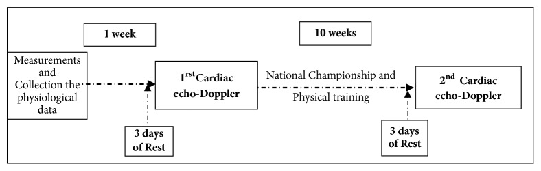
Methods and patients: We conducted a prospective study that included Beninese soccer referees. The study of the geometry and function of the left ventricle (LV) was made at the beginning and end of the national Division 1 championship, which was held during the course of 10 weeks.
Results: There were 37 referees included in this study; 20 at the national level (G1: 27.8 ± 6.6 years) and 17 at the international level (G2: 32.1 ± 6.4 years). Dimensions of the LV were normal for all the referees. At the beginning of the championship, 51.3% of the referees had a normal LV geometry, 37.8% had concentric remodelling, 2.7% had concentric hypertrophy, and 8.1% had eccentric hypertrophy. In the group of referees with normal LV geometry, a modification in concentric remodelling at the end of the championship was seen in 30% of the referees in G1, 33.3% of the referees in G2, and 31.6% of the whole sample. In the group of subjects who presented concentric LV remodelling, a modification in the normal geometry was observed in 37.5% of those in G1, in 0% of those in G2, and in 21.4% of the whole sample. The cases of LV hypertrophy showed no change regardless of the group considered. An LV ejection fraction of more than 50% and an E/E' ratio less than 8 were found in all referees.
Conclusion: All the referees studied had normal cardiac morphology and function. The intensity of the physical load was insufficient to impact this morphology.

 求助内容:
求助内容: 应助结果提醒方式:


