{"title":"Phenylalanine intercalation parameters for liquid-disordered phase domains - a membrane model study.","authors":"Paulina Adamczewski, Valeria Tsoukanova","doi":"10.1186/s13628-018-0047-z","DOIUrl":null,"url":null,"abstract":"<p><strong>Background: </strong>Propensity of phenylalanine (Phe) for nonpolar environments drives its intercalation into phospholipid membranes, which has been implicated in metabolic and neurological disorders. The knowledge of Phe intercalation parameters can be instrumental in understanding various membrane processes triggered by interactions with Phe, in particular the early events leading to the formation of nucleation/docking sites for the self-assembly of Phe amyloid fibrils at the membrane surface.</p><p><strong>Results: </strong>In this study, we used monolayers of phosphatidylethanolamine (DPPE) and phosphatidylcholine (DPPC) to mimic the membrane outer leaflet. Its initial interaction with Phe was modeled by injecting Phe into the aqueous phase underneath the monolayer. Constant pressure insertion assays augmented with epifluorescence microscopy imaging were used to monitor Phe intercalation. Our primary goal was to determine the Phe intercalation area, <i>A</i> <sub>Phe</sub>. Two values were obtained for <i>A</i> <sub>Phe</sub>, 33 ± 2 and 48 ± 3 Å<sup>2</sup>.</p><p><strong>Conclusions: </strong>Phe appeared to discriminate between DPPE and DPPC packing, and use two modes of intercalation. The area of <i>A</i> <sub>Phe</sub> 33 ± 2 Å<sup>2</sup> is consistent with a Phe monomer intercalating into membrane by inserting the phenyl ring nearly normal to the membrane surface. This mode has been found to operate in DPPE membranes. For DPPC membranes however, the value of <i>A</i> <sub>Phe</sub> = 48 ± 3 Å<sup>2</sup> suggests that, from saline, Phe tends to intercalate as a larger species plausibly dragging along a counterion, Na<sup>+</sup>, in a Na<sup>+</sup>(Phe) complex.</p>","PeriodicalId":9045,"journal":{"name":"BMC Biophysics","volume":"11 ","pages":"6"},"PeriodicalIF":0.0000,"publicationDate":"2018-11-15","publicationTypes":"Journal Article","fieldsOfStudy":null,"isOpenAccess":false,"openAccessPdf":"https://sci-hub-pdf.com/10.1186/s13628-018-0047-z","citationCount":"4","resultStr":null,"platform":"Semanticscholar","paperid":null,"PeriodicalName":"BMC Biophysics","FirstCategoryId":"1085","ListUrlMain":"https://doi.org/10.1186/s13628-018-0047-z","RegionNum":0,"RegionCategory":null,"ArticlePicture":[],"TitleCN":null,"AbstractTextCN":null,"PMCID":null,"EPubDate":"","PubModel":"","JCR":"Q1","JCRName":"Biochemistry, Genetics and Molecular Biology","Score":null,"Total":0}
引用次数: 4
Abstract
Background: Propensity of phenylalanine (Phe) for nonpolar environments drives its intercalation into phospholipid membranes, which has been implicated in metabolic and neurological disorders. The knowledge of Phe intercalation parameters can be instrumental in understanding various membrane processes triggered by interactions with Phe, in particular the early events leading to the formation of nucleation/docking sites for the self-assembly of Phe amyloid fibrils at the membrane surface.
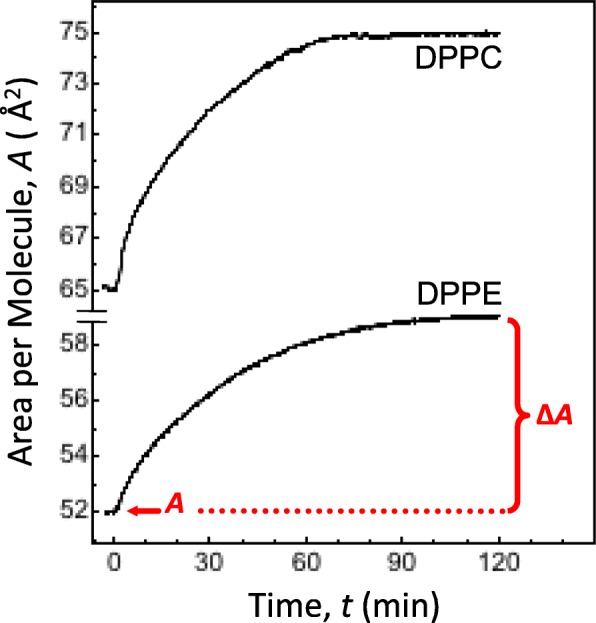
Results: In this study, we used monolayers of phosphatidylethanolamine (DPPE) and phosphatidylcholine (DPPC) to mimic the membrane outer leaflet. Its initial interaction with Phe was modeled by injecting Phe into the aqueous phase underneath the monolayer. Constant pressure insertion assays augmented with epifluorescence microscopy imaging were used to monitor Phe intercalation. Our primary goal was to determine the Phe intercalation area, APhe. Two values were obtained for APhe, 33 ± 2 and 48 ± 3 Å2.
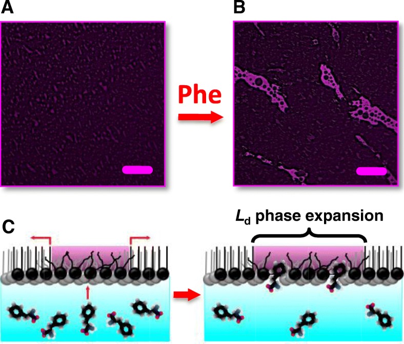
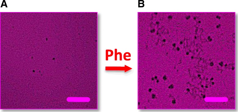
Conclusions: Phe appeared to discriminate between DPPE and DPPC packing, and use two modes of intercalation. The area of APhe 33 ± 2 Å2 is consistent with a Phe monomer intercalating into membrane by inserting the phenyl ring nearly normal to the membrane surface. This mode has been found to operate in DPPE membranes. For DPPC membranes however, the value of APhe = 48 ± 3 Å2 suggests that, from saline, Phe tends to intercalate as a larger species plausibly dragging along a counterion, Na+, in a Na+(Phe) complex.

 求助内容:
求助内容: 应助结果提醒方式:


