Bo Xie, Xinyu Liu, Jie Yang, Jinke Cheng, Jianmin Gu, Song Xue
{"title":"PIAS1 protects against myocardial ischemia-reperfusion injury by stimulating PPARγ SUMOylation.","authors":"Bo Xie, Xinyu Liu, Jie Yang, Jinke Cheng, Jianmin Gu, Song Xue","doi":"10.1186/s12860-018-0176-x","DOIUrl":null,"url":null,"abstract":"<p><strong>Background: </strong>Myocardial ischemia-reperfusion injury (IRI) has become one of the most serious complications after reperfusion therapy in patients with acute myocardial infarction. Small ubiquitin-like modification (SUMOylation) is a reversible process, including SUMO E1-, E2-, and E3-mediated SUMOylation and SUMO-specific protease-mediated deSUMOylation, with the latter having been shown to play a vital role in myocardial IRI previously. However, little is known about the function and regulation of SUMO E3 ligases in myocardial IRI.</p><p><strong>Results: </strong>In this study, we found dramatically decreased expression of PIAS1 after ischemia/reperfusion (I/R) in mouse myocardium and H9C2 cells. PIAS1 deficiency aggravated apoptosis and inflammation of cardiomyocytes via activating the NF-κB pathway after I/R. Mechanistically, we identified PIAS1 as a specific E3 ligase for PPARγ SUMOylation. Moreover, H9C2 cells treated with hypoxia/reoxygenation (H/R) displayed reduced PPARγ SUMOylation as a result of down-regulated PIAS1, and act an anti-apoptotic and anti-inflammatory function through repressing NF-κB activity. Finally, overexpression of PIAS1 in H9C2 cells could remarkably ameliorate I/R injury.</p><p><strong>Conclusions: </strong>Collectively, our findings demonstrate the crucial role of PIAS1-mediated PPARγ SUMOylation in protecting against myocardial IRI.</p>","PeriodicalId":9051,"journal":{"name":"BMC Cell Biology","volume":" ","pages":"24"},"PeriodicalIF":0.0000,"publicationDate":"2018-11-12","publicationTypes":"Journal Article","fieldsOfStudy":null,"isOpenAccess":false,"openAccessPdf":"https://sci-hub-pdf.com/10.1186/s12860-018-0176-x","citationCount":"27","resultStr":null,"platform":"Semanticscholar","paperid":null,"PeriodicalName":"BMC Cell Biology","FirstCategoryId":"1085","ListUrlMain":"https://doi.org/10.1186/s12860-018-0176-x","RegionNum":0,"RegionCategory":null,"ArticlePicture":[],"TitleCN":null,"AbstractTextCN":null,"PMCID":null,"EPubDate":"","PubModel":"","JCR":"Q1","JCRName":"Biochemistry, Genetics and Molecular Biology","Score":null,"Total":0}
引用次数: 27
Abstract
Background: Myocardial ischemia-reperfusion injury (IRI) has become one of the most serious complications after reperfusion therapy in patients with acute myocardial infarction. Small ubiquitin-like modification (SUMOylation) is a reversible process, including SUMO E1-, E2-, and E3-mediated SUMOylation and SUMO-specific protease-mediated deSUMOylation, with the latter having been shown to play a vital role in myocardial IRI previously. However, little is known about the function and regulation of SUMO E3 ligases in myocardial IRI.
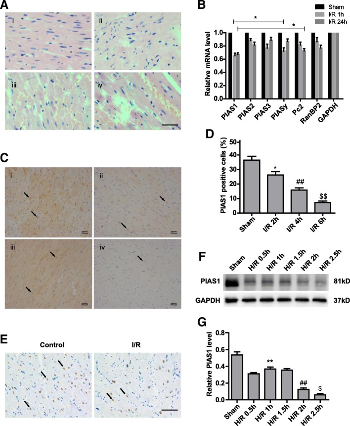
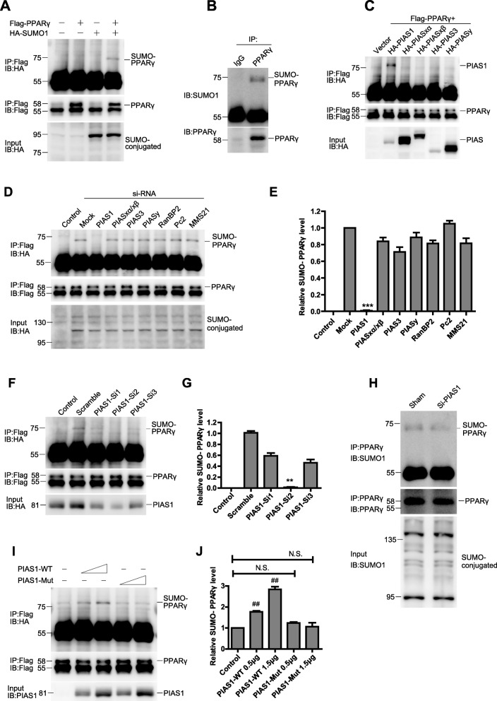
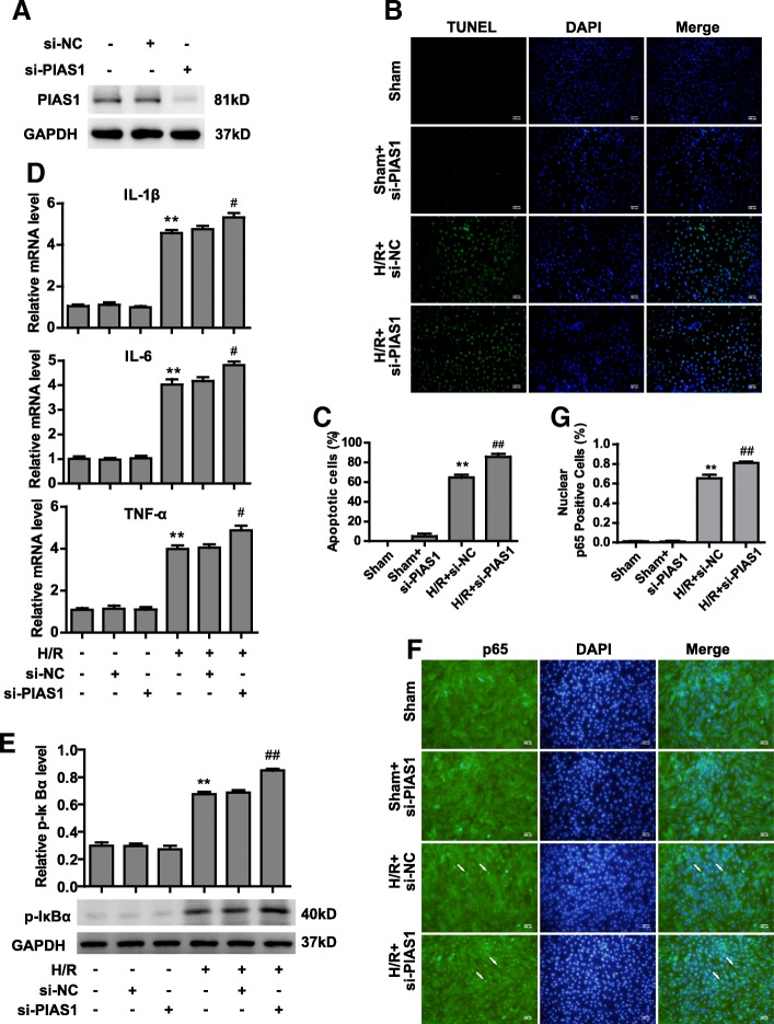
Results: In this study, we found dramatically decreased expression of PIAS1 after ischemia/reperfusion (I/R) in mouse myocardium and H9C2 cells. PIAS1 deficiency aggravated apoptosis and inflammation of cardiomyocytes via activating the NF-κB pathway after I/R. Mechanistically, we identified PIAS1 as a specific E3 ligase for PPARγ SUMOylation. Moreover, H9C2 cells treated with hypoxia/reoxygenation (H/R) displayed reduced PPARγ SUMOylation as a result of down-regulated PIAS1, and act an anti-apoptotic and anti-inflammatory function through repressing NF-κB activity. Finally, overexpression of PIAS1 in H9C2 cells could remarkably ameliorate I/R injury.
Conclusions: Collectively, our findings demonstrate the crucial role of PIAS1-mediated PPARγ SUMOylation in protecting against myocardial IRI.
期刊介绍:
BMC Molecular and Cell Biology, formerly known as BMC Cell Biology, is an open access journal that considers articles on all aspects of both eukaryotic and prokaryotic cell and molecular biology, including structural and functional cell biology, DNA and RNA in a cellular context and biochemistry, as well as research using both the experimental and theoretical aspects of physics to study biological processes and investigations into the structure of biological macromolecules.
 求助内容:
求助内容: 应助结果提醒方式:


