{"title":"MicroRNA-32 Regulates Development and Progression of Hepatocellular Carcinoma by Targeting ADAMTS9 and Affects Its Prognosis.","authors":"Shengmian Li, Tingting Li, Xiaoming Li, Yue Yao, Xiaojia Jiang, Lianmei Zhao, Wei Guo","doi":"10.12659/MSMBR.910522","DOIUrl":null,"url":null,"abstract":"<p><p>BACKGROUND MicroRNA-32 (miR-32) induces cell proliferation and metastasis in hepatocellular carcinoma (HCC), but the detailed mechanisms of miR-32 in regulating oncogenesis and development of HCC have not been clarified. The aim of this study was to investigate the effects of miR-32 on HCC and its clinical pathological significance, as well as to determine the functional connection between miR-32 and ADAMTS9 in HCC. MATERIAL AND METHODS Quantitative RT-PCR was used to assess the expression levels of miR-32 in HCC tissues, adjacent non-cancerous tissues, and liver cancer cell lines. In vitro cell proliferation, migration, and invasion assays were performed to confirm the biological functions of miR-32. Quantitative RT-PCR, Western blot analysis, and luciferase reporter assays were used to evaluate the role of miR-32 in the regulation of ADAMTS9. RESULTS miR-32 was highly expressed in HCC tissues compared with corresponding adjacent non-cancerous tissues. Over-expression of miR-32 was also found in 3 human liver cancer cell lines: SMMC-7721, Huh7, and HepG2. Moreover, increasing expression of miR-32 in HCC tissues was related to shorter overall survival. In vitro over-expression of miR-32 promoted cell proliferation, migration, and invasion; however, the under-expression of miR-32 revealed the opposite effects. Dual-luciferase reporter assay indicated that miR-32 can directly bind to the 3'-UTR of ADAMTS9. Western blot analysis showed that over-expression of miR-32 decreased expression of ADAMTS9 protein. Rescue tests further verified the connection between miR-32 and ADAMTS9. CONCLUSIONS Our data indicate that miR-32 accelerates progression in HCC by targeting ADAMTS9, and the abnormal expression of miR-32 is correlated with prognosis and could become a potential therapeutic target.</p>","PeriodicalId":18491,"journal":{"name":"Medical Science Monitor Basic Research","volume":"24 ","pages":"177-187"},"PeriodicalIF":2.0000,"publicationDate":"2018-11-05","publicationTypes":"Journal Article","fieldsOfStudy":null,"isOpenAccess":false,"openAccessPdf":"https://ftp.ncbi.nlm.nih.gov/pub/pmc/oa_pdf/93/07/medscimonitbasicres-24-177.PMC6237041.pdf","citationCount":"9","resultStr":null,"platform":"Semanticscholar","paperid":null,"PeriodicalName":"Medical Science Monitor Basic Research","FirstCategoryId":"1085","ListUrlMain":"https://doi.org/10.12659/MSMBR.910522","RegionNum":0,"RegionCategory":null,"ArticlePicture":[],"TitleCN":null,"AbstractTextCN":null,"PMCID":null,"EPubDate":"","PubModel":"","JCR":"Q3","JCRName":"MEDICINE, RESEARCH & EXPERIMENTAL","Score":null,"Total":0}
引用次数: 9
Abstract
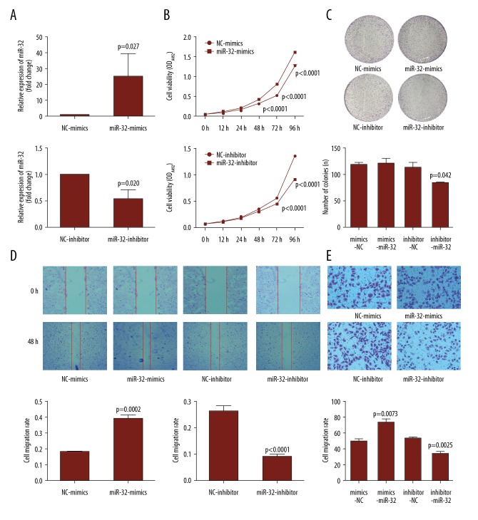
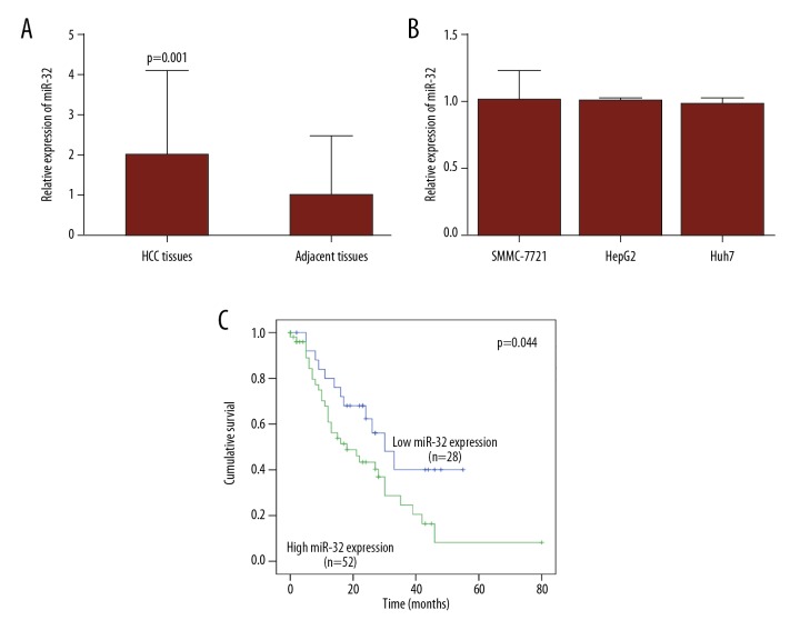
BACKGROUND MicroRNA-32 (miR-32) induces cell proliferation and metastasis in hepatocellular carcinoma (HCC), but the detailed mechanisms of miR-32 in regulating oncogenesis and development of HCC have not been clarified. The aim of this study was to investigate the effects of miR-32 on HCC and its clinical pathological significance, as well as to determine the functional connection between miR-32 and ADAMTS9 in HCC. MATERIAL AND METHODS Quantitative RT-PCR was used to assess the expression levels of miR-32 in HCC tissues, adjacent non-cancerous tissues, and liver cancer cell lines. In vitro cell proliferation, migration, and invasion assays were performed to confirm the biological functions of miR-32. Quantitative RT-PCR, Western blot analysis, and luciferase reporter assays were used to evaluate the role of miR-32 in the regulation of ADAMTS9. RESULTS miR-32 was highly expressed in HCC tissues compared with corresponding adjacent non-cancerous tissues. Over-expression of miR-32 was also found in 3 human liver cancer cell lines: SMMC-7721, Huh7, and HepG2. Moreover, increasing expression of miR-32 in HCC tissues was related to shorter overall survival. In vitro over-expression of miR-32 promoted cell proliferation, migration, and invasion; however, the under-expression of miR-32 revealed the opposite effects. Dual-luciferase reporter assay indicated that miR-32 can directly bind to the 3'-UTR of ADAMTS9. Western blot analysis showed that over-expression of miR-32 decreased expression of ADAMTS9 protein. Rescue tests further verified the connection between miR-32 and ADAMTS9. CONCLUSIONS Our data indicate that miR-32 accelerates progression in HCC by targeting ADAMTS9, and the abnormal expression of miR-32 is correlated with prognosis and could become a potential therapeutic target.


 求助内容:
求助内容: 应助结果提醒方式:


