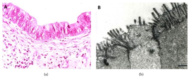
{"title":"HER2 Basolateral versus Circumferential IHC Expression Is Dependent on Polarity and Differentiation of Epithelial Cells in Gastric/GE Adenocarcinoma.","authors":"Shahid Pervez, Sidra Arshad, Brooj Abro","doi":"10.1155/2018/6246493","DOIUrl":null,"url":null,"abstract":"<p><strong>Aim: </strong>Antigenic expression in epithelial cells can be heterogeneous which may pose a problem in immunohistochemical (IHC) analysis of tumor markers, in particular, predictive markers like HER2. Studies have shown that epithelial cells have distinct apical and basolateral domains which are separated by tight junctions. The cell membrane in these two domains has a different composition of macromolecules and hence can have variable antigen expression on immunohistochemistry. In our study, we aimed to investigate this phenomenon of basolateral versus circumferential IHC staining of HER2 in gastric/GE adenocarcinoma.</p><p><strong>Methods: </strong>We selected 45 cases of gastric/GE adenocarcinoma and evaluated equal number of specimens (15 each) showing well-differentiated, moderately differentiated, and poorly differentiated morphology. All cases had 3+ HER2 score as per CAP guidelines. HER2-membrane staining pattern in all specimens was analyzed.</p><p><strong>Results: </strong>Cases with well-differentiated morphology showed only basolateral or lateral membrane staining in most cases. Poorly differentiated adenocarcinoma samples showed circumferential staining (both basolateral and luminal) in all cases with highly significant p value. Mixed staining pattern was observed in moderately differentiated cases. Diffuse expression of E-cadherin in well-differentiated adenocarcinoma and loss in poorly differentiated tumors were also statistically significant.</p><p><strong>Conclusion: </strong>These findings suggest that HER2 in gastric epithelium has a polarized distribution which is maintained by the fence function of tight junctions. With progression to high grade cancer, the glandular structural differentiation in gastric mucosa is lost, along with disruption of tight junctions. This leads to loss of cell polarity and migration of antigens across the membrane.</p>","PeriodicalId":89212,"journal":{"name":"Pathology research international","volume":"2018 ","pages":"6246493"},"PeriodicalIF":0.0000,"publicationDate":"2018-07-24","publicationTypes":"Journal Article","fieldsOfStudy":null,"isOpenAccess":false,"openAccessPdf":"https://sci-hub-pdf.com/10.1155/2018/6246493","citationCount":"1","resultStr":null,"platform":"Semanticscholar","paperid":null,"PeriodicalName":"Pathology research international","FirstCategoryId":"1085","ListUrlMain":"https://doi.org/10.1155/2018/6246493","RegionNum":0,"RegionCategory":null,"ArticlePicture":[],"TitleCN":null,"AbstractTextCN":null,"PMCID":null,"EPubDate":"2018/1/1 0:00:00","PubModel":"eCollection","JCR":"","JCRName":"","Score":null,"Total":0}
引用次数: 1
Abstract
Aim: Antigenic expression in epithelial cells can be heterogeneous which may pose a problem in immunohistochemical (IHC) analysis of tumor markers, in particular, predictive markers like HER2. Studies have shown that epithelial cells have distinct apical and basolateral domains which are separated by tight junctions. The cell membrane in these two domains has a different composition of macromolecules and hence can have variable antigen expression on immunohistochemistry. In our study, we aimed to investigate this phenomenon of basolateral versus circumferential IHC staining of HER2 in gastric/GE adenocarcinoma.
Methods: We selected 45 cases of gastric/GE adenocarcinoma and evaluated equal number of specimens (15 each) showing well-differentiated, moderately differentiated, and poorly differentiated morphology. All cases had 3+ HER2 score as per CAP guidelines. HER2-membrane staining pattern in all specimens was analyzed.
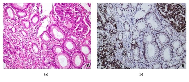
Results: Cases with well-differentiated morphology showed only basolateral or lateral membrane staining in most cases. Poorly differentiated adenocarcinoma samples showed circumferential staining (both basolateral and luminal) in all cases with highly significant p value. Mixed staining pattern was observed in moderately differentiated cases. Diffuse expression of E-cadherin in well-differentiated adenocarcinoma and loss in poorly differentiated tumors were also statistically significant.
Conclusion: These findings suggest that HER2 in gastric epithelium has a polarized distribution which is maintained by the fence function of tight junctions. With progression to high grade cancer, the glandular structural differentiation in gastric mucosa is lost, along with disruption of tight junctions. This leads to loss of cell polarity and migration of antigens across the membrane.


 求助内容:
求助内容: 应助结果提醒方式:


