David Tes, Ahmed Aber, Mohsin Zafar, Luke Horton, Audrey Fotouhi, Qiuyun Xu, Ali Moiin, Andrew D Thompson, Tatiana Cristina Moraes Pinto Blumetti, Steven Daveluy, Wei Chen, Mohammadreza Nasiriavanaki
{"title":"Granular Cell Tumor Imaging Using Optical Coherence Tomography.","authors":"David Tes, Ahmed Aber, Mohsin Zafar, Luke Horton, Audrey Fotouhi, Qiuyun Xu, Ali Moiin, Andrew D Thompson, Tatiana Cristina Moraes Pinto Blumetti, Steven Daveluy, Wei Chen, Mohammadreza Nasiriavanaki","doi":"10.1177/1179597218790250","DOIUrl":null,"url":null,"abstract":"<p><strong>Background: </strong>Granular cell tumor (GCT) is a relatively uncommon tumor that may affect the skin. The tumor can develop anywhere on the body, although it is predominately seen in oral cavities and in the head and neck regions. Here, we present the results of optical coherence tomography (OCT) imaging of a large GCT located on the abdomen of a patient. We also present an analytical method to differentiate between healthy tissue and GCT tissues.</p><p><strong>Materials and methods: </strong>A multibeam, Fourier domain, swept source OCT was used for imaging. The OCT had a central wavelength of 1305 ± 15 nm and lateral and axial resolutions of 7.5 and 10 µm, respectively. Qualitative and quantitative analyses of the tumor and healthy skin are reported.</p><p><strong>Results: </strong>Abrupt changes in architectures of the dermal and epidermal layers in the GCT lesion were observed. These architectural changes were not observed in healthy skin.</p><p><strong>Discussion: </strong>To quantitatively differentiate healthy skin from tumor regions, an optical attenuation coefficient analysis based on single-scattering formulation was performed. The methodology introduced here could have the capability to delineate boundaries of a tumor prior to surgical excision.</p>","PeriodicalId":42484,"journal":{"name":"Biomedical Engineering and Computational Biology","volume":"9 ","pages":"1179597218790250"},"PeriodicalIF":3.1000,"publicationDate":"2018-08-02","publicationTypes":"Journal Article","fieldsOfStudy":null,"isOpenAccess":false,"openAccessPdf":"https://ftp.ncbi.nlm.nih.gov/pub/pmc/oa_pdf/5b/e2/10.1177_1179597218790250.PMC6088518.pdf","citationCount":"0","resultStr":null,"platform":"Semanticscholar","paperid":null,"PeriodicalName":"Biomedical Engineering and Computational Biology","FirstCategoryId":"1085","ListUrlMain":"https://doi.org/10.1177/1179597218790250","RegionNum":0,"RegionCategory":null,"ArticlePicture":[],"TitleCN":null,"AbstractTextCN":null,"PMCID":null,"EPubDate":"2018/1/1 0:00:00","PubModel":"eCollection","JCR":"Q3","JCRName":"ENGINEERING, BIOMEDICAL","Score":null,"Total":0}
引用次数: 0
Abstract
Background: Granular cell tumor (GCT) is a relatively uncommon tumor that may affect the skin. The tumor can develop anywhere on the body, although it is predominately seen in oral cavities and in the head and neck regions. Here, we present the results of optical coherence tomography (OCT) imaging of a large GCT located on the abdomen of a patient. We also present an analytical method to differentiate between healthy tissue and GCT tissues.
Materials and methods: A multibeam, Fourier domain, swept source OCT was used for imaging. The OCT had a central wavelength of 1305 ± 15 nm and lateral and axial resolutions of 7.5 and 10 µm, respectively. Qualitative and quantitative analyses of the tumor and healthy skin are reported.
Results: Abrupt changes in architectures of the dermal and epidermal layers in the GCT lesion were observed. These architectural changes were not observed in healthy skin.
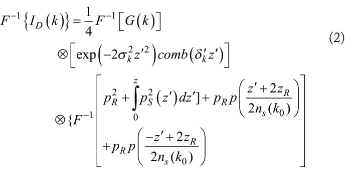
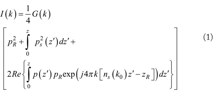
Discussion: To quantitatively differentiate healthy skin from tumor regions, an optical attenuation coefficient analysis based on single-scattering formulation was performed. The methodology introduced here could have the capability to delineate boundaries of a tumor prior to surgical excision.

 求助内容:
求助内容: 应助结果提醒方式:


