Jae Hwi Sung, Ahran Song, Taegun Park, Eunyoung Kim, Seunggwan Lee
{"title":"The Different Expression Patterns of HSP22, a Late Embryogenesis Abundant-like Protein, in Hypertrophic H9C2 Cells Induced by NaCl and Angiotensin II.","authors":"Jae Hwi Sung, Ahran Song, Taegun Park, Eunyoung Kim, Seunggwan Lee","doi":"10.5049/EBP.2018.16.1.1","DOIUrl":null,"url":null,"abstract":"<p><strong>Background: </strong>High-NaCl diet is a contributing factor for cardiac hypertrophy. The role of HSP22 as a protective protein during cardiac hypertrophy due to hypernatremia is unclear. Accordingly, this study aimed to establish a cellular hypernatremic H9C2 model and to compare the expression of HSP22 in Ca<sup>2+</sup> homeostasis between a high-NaCl and angiotensin II-induced hypertrophic cellular H9C2 model.</p><p><strong>Methods: </strong>Real-time PCR was performed to compare the mRNA expression. Flow cytometry and confocal microscopy were used to analyze the cells.</p><p><strong>Results: </strong>The addition of 30 mM NaCl for 48 h was the most effective condition for the induction of hypertrophic H9C2 cells (termed the <i>in vitro</i> hypernatremic model). Cardiac cellular hypertrophy was induced with 30 mM NaCl and 1 µM angiotensin II for 48 h, without causing abnormal morphological changes or cytotoxicity of the culture conditions. HSP22 contains a similar domain to that found in the consensus sequences of the late embryogenesis abundant protein group 3 from <i>Artemia</i>. The expression of HSP22 gradually decreased in the <i>in vitro</i> hypernatremic model. In contrast to the <i>in vitro</i> hypernatremic model, HSP22 increased after exposure to angiotensin II for 48 h. Intracellular Ca<sup>2+</sup> decreased in the angiotensin II model and further decreased in the <i>in vitro</i> hypernatremic model. Impaired intracellular Ca<sup>2+</sup> homeostasis was more evident in the <i>in vitro</i> hypernatremic model.</p><p><strong>Conclusion: </strong>The results showed that NaCl significantly decreased HSP22. Decreased HSP22, due to the hypernatremic condition, affected the Ca<sup>2+</sup> homeostasis in the H9C2 cells. Therefore, hypernatremia induces cellular hypertrophy via impaired Ca<sup>2+</sup> homeostasis. The additional mechanisms of HSP22 need to be explored further.</p>","PeriodicalId":35352,"journal":{"name":"Electrolyte and Blood Pressure","volume":"16 1","pages":"1-10"},"PeriodicalIF":0.0000,"publicationDate":"2018-06-01","publicationTypes":"Journal Article","fieldsOfStudy":null,"isOpenAccess":false,"openAccessPdf":"https://sci-hub-pdf.com/10.5049/EBP.2018.16.1.1","citationCount":"2","resultStr":null,"platform":"Semanticscholar","paperid":null,"PeriodicalName":"Electrolyte and Blood Pressure","FirstCategoryId":"1085","ListUrlMain":"https://doi.org/10.5049/EBP.2018.16.1.1","RegionNum":0,"RegionCategory":null,"ArticlePicture":[],"TitleCN":null,"AbstractTextCN":null,"PMCID":null,"EPubDate":"2018/6/30 0:00:00","PubModel":"Epub","JCR":"Q3","JCRName":"Medicine","Score":null,"Total":0}
引用次数: 2
Abstract
Background: High-NaCl diet is a contributing factor for cardiac hypertrophy. The role of HSP22 as a protective protein during cardiac hypertrophy due to hypernatremia is unclear. Accordingly, this study aimed to establish a cellular hypernatremic H9C2 model and to compare the expression of HSP22 in Ca2+ homeostasis between a high-NaCl and angiotensin II-induced hypertrophic cellular H9C2 model.
Methods: Real-time PCR was performed to compare the mRNA expression. Flow cytometry and confocal microscopy were used to analyze the cells.
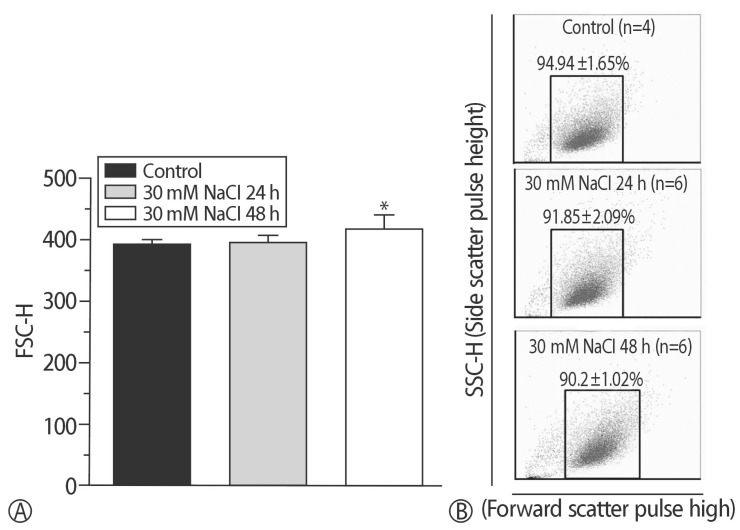
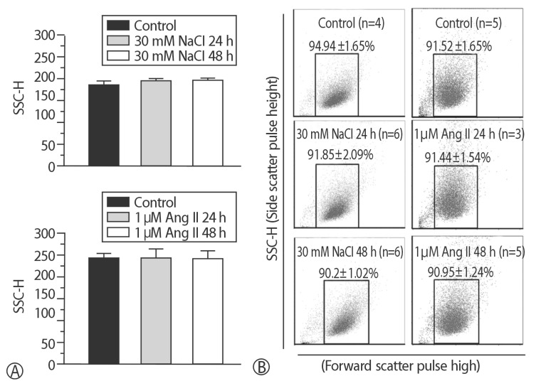
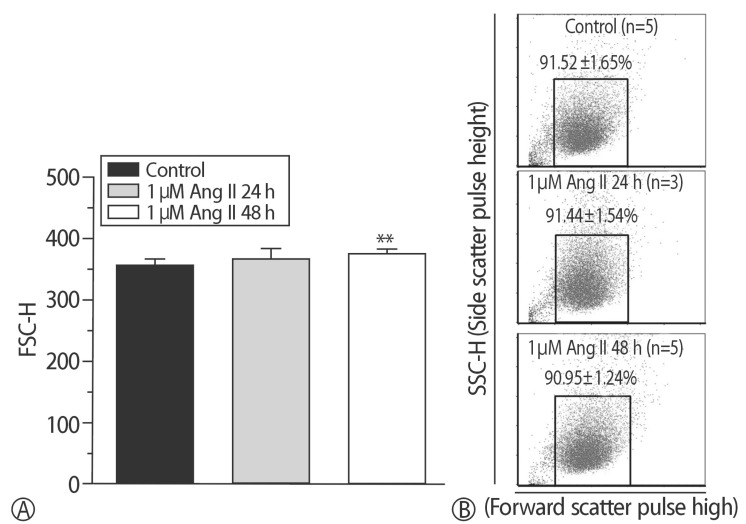
Results: The addition of 30 mM NaCl for 48 h was the most effective condition for the induction of hypertrophic H9C2 cells (termed the in vitro hypernatremic model). Cardiac cellular hypertrophy was induced with 30 mM NaCl and 1 µM angiotensin II for 48 h, without causing abnormal morphological changes or cytotoxicity of the culture conditions. HSP22 contains a similar domain to that found in the consensus sequences of the late embryogenesis abundant protein group 3 from Artemia. The expression of HSP22 gradually decreased in the in vitro hypernatremic model. In contrast to the in vitro hypernatremic model, HSP22 increased after exposure to angiotensin II for 48 h. Intracellular Ca2+ decreased in the angiotensin II model and further decreased in the in vitro hypernatremic model. Impaired intracellular Ca2+ homeostasis was more evident in the in vitro hypernatremic model.
Conclusion: The results showed that NaCl significantly decreased HSP22. Decreased HSP22, due to the hypernatremic condition, affected the Ca2+ homeostasis in the H9C2 cells. Therefore, hypernatremia induces cellular hypertrophy via impaired Ca2+ homeostasis. The additional mechanisms of HSP22 need to be explored further.
背景:高盐饮食是导致心脏肥厚的一个因素。在高钠血症引起的心脏肥厚过程中,HSP22作为一种保护蛋白的作用尚不清楚。因此,本研究旨在建立细胞高钠血症H9C2模型,并比较高nacl和血管紧张素ii诱导的增生性细胞H9C2模型中HSP22在Ca2+稳态中的表达。方法:采用Real-time PCR法比较mRNA的表达情况。采用流式细胞术和共聚焦显微镜对细胞进行分析。结果:30 mM NaCl作用48 h是诱导H9C2细胞增生性的最有效条件(称为体外高钠血症模型)。用30 mM NaCl和1µM血管紧张素II诱导心肌细胞肥大48 h,培养条件未出现异常形态学变化和细胞毒性。HSP22包含一个类似的结构域,该结构域与在Artemia胚胎发育晚期丰富蛋白组3的共识序列中发现的结构域相似。在体外高钠血症模型中,HSP22的表达逐渐降低。与体外高钠血症模型相比,HSP22在暴露于血管紧张素II 48小时后升高。血管紧张素II模型中细胞内Ca2+降低,体外高钠血症模型中进一步降低。在体外高钠血症模型中,细胞内Ca2+稳态受损更为明显。结论:NaCl能显著降低HSP22。高钠血症导致HSP22降低,影响H9C2细胞内Ca2+稳态。因此,高钠血症通过Ca2+稳态受损诱导细胞肥大。HSP22的其他机制有待进一步探索。

 求助内容:
求助内容: 应助结果提醒方式:


