Yassir A Shuaib, Eltahir A G Khalil, Ulrich E Schaible, Lothar H Wieler, Mohammed A M Bakheit, Saad E Mohamed-Noor, Mohamed A Abdalla, Susanne Homolka, Sönke Andres, Doris Hillemann, Knut Lonnroth, Elvira Richter, Stefan Niemann, Katharina Kranzer
{"title":"Smear Microscopy for Diagnosis of Pulmonary Tuberculosis in Eastern Sudan.","authors":"Yassir A Shuaib, Eltahir A G Khalil, Ulrich E Schaible, Lothar H Wieler, Mohammed A M Bakheit, Saad E Mohamed-Noor, Mohamed A Abdalla, Susanne Homolka, Sönke Andres, Doris Hillemann, Knut Lonnroth, Elvira Richter, Stefan Niemann, Katharina Kranzer","doi":"10.1155/2018/8038137","DOIUrl":null,"url":null,"abstract":"<p><strong>Background: </strong>In Sudan, tuberculosis diagnosis largely relies on clinical symptoms and smear microscopy as in many other low- and middle-income countries. The aim of this study was to investigate the positive predictive value of a positive sputum smear in patients investigated for pulmonary tuberculosis in Eastern Sudan.</p><p><strong>Methods: </strong>Two sputum samples from patients presenting with symptoms suggestive of tuberculosis were investigated using direct Ziehl-Neelsen (ZN) staining and light microscopy between June to October 2014 and January to July 2016. If one of the samples was smear positive, both samples were pooled, stored at -20°C, and sent to the National Reference Laboratory (NRL), Germany. Following decontamination, samples underwent repeat microscopy and culture. Culture negative/contaminated samples were investigated using polymerase chain reaction (PCR).</p><p><strong>Results: </strong>A total of 383 samples were investigated. Repeat microscopy categorized 123 (32.1%) as negative, among which 31 were culture positive. This increased to 80 when PCR and culture results were considered together. A total of 196 samples were culture positive, of which 171 (87.3%), 14 (7.1%), and 11 (5.6%) were <i>M. tuberculosis, M. intracellulare</i>, and mixed species. Overall, 15.6% (57/365) of the samples had no evidence of <i>M. tuberculosis</i>, resulting in a positive predictive value of 84.4%.</p><p><strong>Conclusions: </strong>There was a discordance between the results of smear microscopy performed at local laboratories in the Sudan and at the NRL, Germany; besides, a considerable number of samples had no evidence of <i>M. tuberculosis</i>. Improved quality control for smear microscopy and more specific diagnostics are crucial to avoid possible overtreatment.</p>","PeriodicalId":30261,"journal":{"name":"Tuberculosis Research and Treatment","volume":"2018 ","pages":"8038137"},"PeriodicalIF":0.0000,"publicationDate":"2018-06-14","publicationTypes":"Journal Article","fieldsOfStudy":null,"isOpenAccess":false,"openAccessPdf":"https://sci-hub-pdf.com/10.1155/2018/8038137","citationCount":"6","resultStr":null,"platform":"Semanticscholar","paperid":null,"PeriodicalName":"Tuberculosis Research and Treatment","FirstCategoryId":"1085","ListUrlMain":"https://doi.org/10.1155/2018/8038137","RegionNum":0,"RegionCategory":null,"ArticlePicture":[],"TitleCN":null,"AbstractTextCN":null,"PMCID":null,"EPubDate":"2018/1/1 0:00:00","PubModel":"eCollection","JCR":"","JCRName":"","Score":null,"Total":0}
引用次数: 6
Abstract
Background: In Sudan, tuberculosis diagnosis largely relies on clinical symptoms and smear microscopy as in many other low- and middle-income countries. The aim of this study was to investigate the positive predictive value of a positive sputum smear in patients investigated for pulmonary tuberculosis in Eastern Sudan.
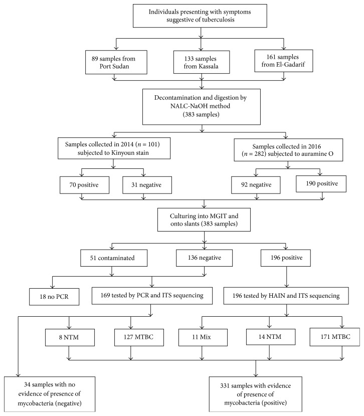
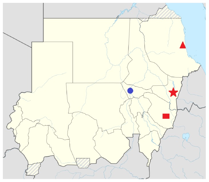
Methods: Two sputum samples from patients presenting with symptoms suggestive of tuberculosis were investigated using direct Ziehl-Neelsen (ZN) staining and light microscopy between June to October 2014 and January to July 2016. If one of the samples was smear positive, both samples were pooled, stored at -20°C, and sent to the National Reference Laboratory (NRL), Germany. Following decontamination, samples underwent repeat microscopy and culture. Culture negative/contaminated samples were investigated using polymerase chain reaction (PCR).
Results: A total of 383 samples were investigated. Repeat microscopy categorized 123 (32.1%) as negative, among which 31 were culture positive. This increased to 80 when PCR and culture results were considered together. A total of 196 samples were culture positive, of which 171 (87.3%), 14 (7.1%), and 11 (5.6%) were M. tuberculosis, M. intracellulare, and mixed species. Overall, 15.6% (57/365) of the samples had no evidence of M. tuberculosis, resulting in a positive predictive value of 84.4%.
Conclusions: There was a discordance between the results of smear microscopy performed at local laboratories in the Sudan and at the NRL, Germany; besides, a considerable number of samples had no evidence of M. tuberculosis. Improved quality control for smear microscopy and more specific diagnostics are crucial to avoid possible overtreatment.

 求助内容:
求助内容: 应助结果提醒方式:


